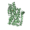
 Movie
Movie Controller

[English] 日本語
 Yorodumi
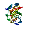
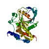
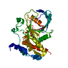
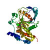
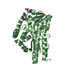
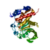
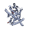
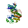
- PDB-3o4s: Crystal Structure of HePTP with a Closed WPD Loop and an Ordered ... -
+ Open data

- Basic information
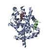
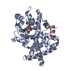
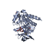
| Entry | Database: PDB / ID: 3o4s | ||||||
|---|---|---|---|---|---|---|---|
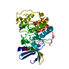
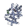
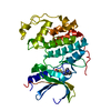
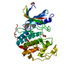
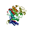
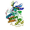
| Title | Crystal Structure of HePTP with a Closed WPD Loop and an Ordered E-Loop | ||||||
 Components Components | Tyrosine-protein phosphatase non-receptor type 7 | ||||||
 Keywords Keywords | HYDROLASE / HEPTP / HUMAN HEMATOPOIETIC TYROSINE PHOSPHATASE CATALYTIC DOMAIN MUTANT / LC-PTP / PTPN7 | ||||||
| Function / homology |  Function and homology information Function and homology informationInterleukin-37 signaling / protein dephosphorylation / non-membrane spanning protein tyrosine phosphatase activity / protein-tyrosine-phosphatase / protein tyrosine phosphatase activity / cytoplasmic side of plasma membrane / Negative regulation of MAPK pathway / mitotic spindle / MAPK cascade / microtubule cytoskeleton ...Interleukin-37 signaling / protein dephosphorylation / non-membrane spanning protein tyrosine phosphatase activity / protein-tyrosine-phosphatase / protein tyrosine phosphatase activity / cytoplasmic side of plasma membrane / Negative regulation of MAPK pathway / mitotic spindle / MAPK cascade / microtubule cytoskeleton / signal transduction / nucleoplasm / cytoplasm / cytosol Similarity search - Function | ||||||
| Biological species |  Homo sapiens (human) Homo sapiens (human) | ||||||
| Method |  X-RAY DIFFRACTION / SYNCHROTRON / FOURIER SYNTHESIS / Resolution: 1.9 Å X-RAY DIFFRACTION / SYNCHROTRON / FOURIER SYNTHESIS / Resolution: 1.9 Å | ||||||
 Authors Authors | Critton, D.A. / Page, R. | ||||||
 Citation Citation | Journal: J.Mol.Biol. / Year: 2011 Title: Visualizing active-site dynamics in single crystals of HePTP: opening of the WPD loop involves coordinated movement of the E loop. Authors: Critton, D.A. / Tautz, L. / Page, R. | ||||||
| History |
|
- Structure visualization
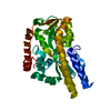
| Structure viewer | Molecule: MolmilJmol/JSmol |
|---|
- Downloads & links
-Download
| PDBx/mmCIF format | 3o4s.cif.gz | 81.9 KB | Display | PDBx/mmCIF format |
|---|---|---|---|---|
| PDB format | pdb3o4s.ent.gz | 59.6 KB | Display | PDB format |
| PDBx/mmJSON format | 3o4s.json.gz | Tree view | PDBx/mmJSON format | |
| Others |  Other downloads Other downloads |
-Validation report
| Arichive directory | https://data.pdbj.org/pub/pdb/validation_reports/o4/3o4sftp://data.pdbj.org/pub/pdb/validation_reports/o4/3o4s | HTTPS FTP |
|---|
-Related structure data
| Related structure data | 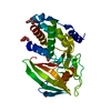 3o4tC  3o4uC 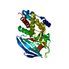 2qdmS S: Starting model for refinement C: citing same article ( |
|---|---|
| Similar structure data |


-Links
 PDBj
PDBj


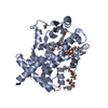
- Assembly
| Deposited unit | 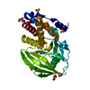
| ||||||||
|---|---|---|---|---|---|---|---|---|---|
| 1 |
| ||||||||
| Unit cell |
|
-Components
| #1: Protein | Mass: 35232.801 Da / Num. of mol.: 1 / Fragment: UNP RESIDUES 65-360 / Mutation: S72D Source method: isolated from a genetically manipulated source Source: (gene. exp.) Homo sapiens (human) / Gene: PTPN7 / Plasmid: PBAD / Production host:  | ||||
|---|---|---|---|---|---|
| #2: Chemical | ChemComp-SO4 /   Mass: 96.063 Da / Num. of mol.: 6 / Source method: obtained synthetically / Formula: SO4 Mass: 96.063 Da / Num. of mol.: 6 / Source method: obtained synthetically / Formula: SO4#3: Chemical | ChemComp-GOL /   Mass: 92.094 Da / Num. of mol.: 5 / Source method: obtained synthetically / Formula: C3H8O3 Mass: 92.094 Da / Num. of mol.: 5 / Source method: obtained synthetically / Formula: C3H8O3#4: Water | ChemComp-HOH / |  Mass: 18.015 Da / Num. of mol.: 277 / Source method: isolated from a natural source / Formula: H2O Mass: 18.015 Da / Num. of mol.: 277 / Source method: isolated from a natural source / Formula: H2O |
-Experimental details
-Experiment
| Experiment | Method: X-RAY DIFFRACTION / Number of used crystals: 1 |
|---|
- Sample preparation
| Crystal | Density Matthews: 2.25 Å3/Da / Density % sol: 45.45 % |
|---|---|
| Crystal grow | Temperature: 277 K / Method: vapor diffusion, sitting drop / pH: 5 Details: 1.7-1.9M ammonium sulfate, pH 5.0, VAPOR DIFFUSION, SITTING DROP, temperature 277K |
-Data collection
| Diffraction | Mean temperature: 100 K |
|---|---|
| Diffraction source | Source: SYNCHROTRON / Site: NSLS  / Beamline: X6A / Wavelength: 1 Å / Beamline: X6A / Wavelength: 1 Å |
| Detector | Type: ADSC QUANTUM 270 / Detector: CCD / Date: Apr 23, 2009 / Details: OXFORD DANFYSIK TOROIDAL FOCUSING MIRROR |
| Radiation | Monochromator: SI(III) CHANNEL CUT MONOCHROMATOR / Protocol: SINGLE WAVELENGTH / Monochromatic (M) / Laue (L): M / Scattering type: x-ray |
| Radiation wavelength | Wavelength: 1 Å / Relative weight: 1 |
| Reflection | Resolution: 1.9→50 Å / Num. all: 25307 / Num. obs: 25241 / % possible obs: 99.7 % / Redundancy: 3.7 % / Biso Wilson estimate: 19.1 Å2 / Rsym value: 0.092 / Net I/σ(I): 13.8 |
| Reflection shell | Resolution: 1.9→1.93 Å / Redundancy: 3.6 % / Mean I/σ(I) obs: 3.5 / Num. unique all: 1246 / Rsym value: 0.512 / % possible all: 99.9 |
- Processing
| Software |
| ||||||||||||||||||||||||||||||||||||||||||||||||||||||||||||||||||||||||||||||||||||||||||
|---|---|---|---|---|---|---|---|---|---|---|---|---|---|---|---|---|---|---|---|---|---|---|---|---|---|---|---|---|---|---|---|---|---|---|---|---|---|---|---|---|---|---|---|---|---|---|---|---|---|---|---|---|---|---|---|---|---|---|---|---|---|---|---|---|---|---|---|---|---|---|---|---|---|---|---|---|---|---|---|---|---|---|---|---|---|---|---|---|---|---|---|
| Refinement | Method to determine structure: FOURIER SYNTHESIS Starting model: PDB ENTRY 2QDM Resolution: 1.9→20 Å / Cor.coef. Fo:Fc: 0.963 / Cor.coef. Fo:Fc free: 0.933 / SU B: 2.98 / SU ML: 0.088 / Cross valid method: THROUGHOUT / ESU R Free: 0.137 / Stereochemistry target values: MAXIMUM LIKELIHOOD Details: HYDROGENS HAVE BEEN ADDED IN THE RIDING POSITIONS RESIDUES AND SIDECHAINS FOR WHICH NO DENSITY WAS OBSERVED WERE NOT MODELED
| ||||||||||||||||||||||||||||||||||||||||||||||||||||||||||||||||||||||||||||||||||||||||||
| Solvent computation | Ion probe radii: 0.8 Å / Shrinkage radii: 0.8 Å / VDW probe radii: 1.2 Å / Solvent model: MASK | ||||||||||||||||||||||||||||||||||||||||||||||||||||||||||||||||||||||||||||||||||||||||||
| Displacement parameters | Biso mean: 21.059 Å2
| ||||||||||||||||||||||||||||||||||||||||||||||||||||||||||||||||||||||||||||||||||||||||||
| Refinement step | Cycle: LAST / Resolution: 1.9→20 Å
| ||||||||||||||||||||||||||||||||||||||||||||||||||||||||||||||||||||||||||||||||||||||||||
| Refine LS restraints |
| ||||||||||||||||||||||||||||||||||||||||||||||||||||||||||||||||||||||||||||||||||||||||||
| LS refinement shell | Resolution: 1.9→1.944 Å / Total num. of bins used: 20
|
