 Movie
Movie Controller

[English] 日本語
 Yorodumi
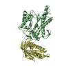
- PDB-3asy: ligand-free structure of uridine kinase from thermus thermophilus HB8 -
+ Open data

- Basic information
| Entry | Database: PDB / ID: 3asy | ||||||
|---|---|---|---|---|---|---|---|
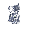
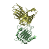
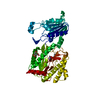
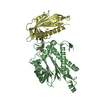
| Title | ligand-free structure of uridine kinase from thermus thermophilus HB8 | ||||||
 Components Components | Uridine kinase | ||||||
 Keywords Keywords | TRANSFERASE / cytidine phosphorylation | ||||||
| Function / homology |  Function and homology information Function and homology informationuridine/cytidine kinase / CTP salvage / uridine kinase activity / cytidine kinase activity / UMP salvage / ATP binding / cytoplasm Similarity search - Function | ||||||
| Biological species |   Thermus thermophilus (bacteria) Thermus thermophilus (bacteria) | ||||||
| Method |  X-RAY DIFFRACTION / SYNCHROTRON / MOLECULAR REPLACEMENT / Resolution: 2.4 Å X-RAY DIFFRACTION / SYNCHROTRON / MOLECULAR REPLACEMENT / Resolution: 2.4 Å | ||||||
 Authors Authors | Tomoike, F. / Nakagawa, N. / Kuramitsu, S. / Masui, R. | ||||||
 Citation Citation | Journal: Biochemistry / Year: 2011 Title: A Single Amino Acid Limits the Substrate Specificity of Thermus thermophilus Uridine-Cytidine Kinase to Cytidine Authors: Tomoike, F. / Nakagawa, N. / Kuramitsu, S. / Masui, R. | ||||||
| History |
|
- Structure visualization
| Structure viewer | Molecule: MolmilJmol/JSmol |
|---|
- Downloads & links
-Download
| PDBx/mmCIF format | 3asy.cif.gz | 90 KB | Display | PDBx/mmCIF format |
|---|---|---|---|---|
| PDB format | pdb3asy.ent.gz | 69.8 KB | Display | PDB format |
| PDBx/mmJSON format | 3asy.json.gz | Tree view | PDBx/mmJSON format | |
| Others |  Other downloads Other downloads |
-Validation report
| Arichive directory | https://data.pdbj.org/pub/pdb/validation_reports/as/3asyftp://data.pdbj.org/pub/pdb/validation_reports/as/3asy | HTTPS FTP |
|---|
-Related structure data


-Links
 PDBj
PDBj- Assembly
| Deposited unit | 
| ||||||||
|---|---|---|---|---|---|---|---|---|---|
| 1 |
| ||||||||
| Unit cell |
|
-Components
| #1: Protein | Mass: 23709.723 Da / Num. of mol.: 2 Source method: isolated from a genetically manipulated source Source: (gene. exp.) Thermus thermophilus (bacteria) / Strain: HB8 / Gene: udk, TTHA0578 / Plasmid: pET11a / Production host: #2: Water | ChemComp-HOH / |  Mass: 18.015 Da / Num. of mol.: 82 / Source method: isolated from a natural source / Formula: H2O Mass: 18.015 Da / Num. of mol.: 82 / Source method: isolated from a natural source / Formula: H2O |
|---|
-Experimental details
-Experiment
| Experiment | Method: X-RAY DIFFRACTION / Number of used crystals: 1 |
|---|
- Sample preparation
| Crystal | Density Matthews: 2.34 Å3/Da / Density % sol: 47.42 % |
|---|---|
| Crystal grow | Temperature: 293 K / Method: vapor diffusion / pH: 6.5 Details: 40% (v/v) isopropanol, 15%(w/v) PEG 8000, 0.1M imidazole, pH 6.5, VAPOR DIFFUSION, temperature 293K |
-Data collection
| Diffraction | Mean temperature: 100 K |
|---|---|
| Diffraction source | Source: SYNCHROTRON / Site: SPring-8  / Beamline: BL26B2 / Wavelength: 1 Å / Beamline: BL26B2 / Wavelength: 1 Å |
| Detector | Type: MARMOSAIC 225 mm CCD / Detector: CCD / Date: Jan 31, 2008 |
| Radiation | Monochromator: transparent diamond double crystal / Protocol: SINGLE WAVELENGTH / Monochromatic (M) / Laue (L): M / Scattering type: x-ray |
| Radiation wavelength | Wavelength: 1 Å / Relative weight: 1 |
| Reflection | Resolution: 2.4→50 Å / Num. all: 18465 / Num. obs: 18465 / Observed criterion σ(F): 0 |
| Reflection shell | Resolution: 2.4→2.49 Å / Rmerge(I) obs: 0.64 / Mean I/σ(I) obs: 6.4 |
- Processing
| Software |
| |||||||||||||||||||||||||
|---|---|---|---|---|---|---|---|---|---|---|---|---|---|---|---|---|---|---|---|---|---|---|---|---|---|---|
| Refinement | Method to determine structure: MOLECULAR REPLACEMENT Starting model: uridine cytidine kinase2 from Homo sapiens Resolution: 2.4→37.8 Å / σ(F): 0
| |||||||||||||||||||||||||
| Solvent computation | Bsol: 27.6311 Å2 | |||||||||||||||||||||||||
| Displacement parameters | Biso mean: 47.0139 Å2
| |||||||||||||||||||||||||
| Refinement step | Cycle: LAST / Resolution: 2.4→37.8 Å
| |||||||||||||||||||||||||
| Refine LS restraints |
| |||||||||||||||||||||||||
| Xplor file |
|
