 Movie
Movie Controller

[English] 日本語
 Yorodumi
- PDB-309d: A DNA DECAMER WITH A STICKY END: THE CRYSTAL STRUCTURE OF D-CGACGATCGT -
+ Open data

- Basic information
| Entry | Database: PDB / ID: 309d | |||||||||||||||||||||
|---|---|---|---|---|---|---|---|---|---|---|---|---|---|---|---|---|---|---|---|---|---|---|
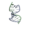
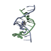
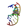
| Title | A DNA DECAMER WITH A STICKY END: THE CRYSTAL STRUCTURE OF D-CGACGATCGT | |||||||||||||||||||||
 Components Components | DNA (5'-D(* Keywords Keywords DNA / B-DNA / DOUBLE HELIX / OVERHANGING BASES / CONTINUOUS HELIX DNA / B-DNA / DOUBLE HELIX / OVERHANGING BASES / CONTINUOUS HELIXFunction / homology | DNA Function and homology information Function and homology informationMethod | X-RAY DIFFRACTION / MOLECULAR REPLACEMENT / Resolution: 2.6 Å  Authors AuthorsQiu, H.X. / Dewan, J.C. / Seeman, N.C. |  CitationJournal: J.Mol.Biol. / Year: 1997 CitationJournal: J.Mol.Biol. / Year: 1997Title: A DNA decamer with a sticky end: the crystal structure of d-CGACGATCGT. Authors: Qiu, H. / Dewan, J.C. / Seeman, N.C. History |
|
- Structure visualization
| Structure viewer | Molecule: MolmilJmol/JSmol |
|---|
- Downloads & links
-Download
| PDBx/mmCIF format | 309d.cif.gz | 20 KB | Display | PDBx/mmCIF format |
|---|---|---|---|---|
| PDB format | pdb309d.ent.gz | 12.7 KB | Display | PDB format |
| PDBx/mmJSON format | 309d.json.gz | Tree view | PDBx/mmJSON format | |
| Others |  Other downloads Other downloads |
-Validation report
| Arichive directory | https://data.pdbj.org/pub/pdb/validation_reports/09/309dftp://data.pdbj.org/pub/pdb/validation_reports/09/309d | HTTPS FTP |
|---|
-Related structure data
| Similar structure data |
|---|


-Links
 PDBj
PDBj


- Assembly
| Deposited unit | 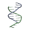
| ||||||||
|---|---|---|---|---|---|---|---|---|---|
| 1 |
| ||||||||
| Unit cell |
|
-Components
| #1: DNA chain | Mass: 3045.004 Da / Num. of mol.: 2 / Source method: obtained synthetically #2: Water | ChemComp-HOH / | Water Mass: 18.015 Da / Num. of mol.: 68 / Source method: isolated from a natural source / Formula: H2O Mass: 18.015 Da / Num. of mol.: 68 / Source method: isolated from a natural source / Formula: H2O |
|---|
-Experimental details
-Experiment
| Experiment | Method: X-RAY DIFFRACTION |
|---|
- Sample preparation
| Crystal | Density Matthews: 2.22 Å3/Da / Density % sol: 44.65 % | |||||||||||||||||||||||||||||||||||
|---|---|---|---|---|---|---|---|---|---|---|---|---|---|---|---|---|---|---|---|---|---|---|---|---|---|---|---|---|---|---|---|---|---|---|---|---|
| Crystal grow | Temperature: 291 K / Method: vapor diffusion, sitting drop / Details: VAPOR DIFFUSION, SITTING DROP, temperature 291K | |||||||||||||||||||||||||||||||||||
| Components of the solutions |
| |||||||||||||||||||||||||||||||||||
| Crystal grow | *PLUS | |||||||||||||||||||||||||||||||||||
| Components of the solutions | *PLUS
|
-Data collection
| Diffraction | Mean temperature: 153 K |
|---|---|
| Diffraction source | Source: ROTATING ANODE / Type: RIGAKU RU200 / Wavelength: 1.5418 |
| Detector | Type: RIGAKU AFC-5R / Detector: DIFFRACTOMETER |
| Radiation | Scattering type: x-ray |
| Radiation wavelength | Wavelength: 1.5418 Å / Relative weight: 1 |
| Reflection | Highest resolution: 2.6 Å / Num. obs: 1768 / Observed criterion σ(I): 2 |
| Reflection shell | Resolution: 2.6→3.6 Å / % possible all: 75 |
- Processing
| Software | Name: NUCLSQ / Classification: refinement | ||||||||||||||||||||||||||||||||||||||||||||||||||||||||||||||||||||||||||||||||||||
|---|---|---|---|---|---|---|---|---|---|---|---|---|---|---|---|---|---|---|---|---|---|---|---|---|---|---|---|---|---|---|---|---|---|---|---|---|---|---|---|---|---|---|---|---|---|---|---|---|---|---|---|---|---|---|---|---|---|---|---|---|---|---|---|---|---|---|---|---|---|---|---|---|---|---|---|---|---|---|---|---|---|---|---|---|---|
| Refinement | Method to determine structure: MOLECULAR REPLACEMENT / Resolution: 2.6→8 Å / σ(F): 2 /
| ||||||||||||||||||||||||||||||||||||||||||||||||||||||||||||||||||||||||||||||||||||
| Refinement step | Cycle: LAST / Resolution: 2.6→8 Å
| ||||||||||||||||||||||||||||||||||||||||||||||||||||||||||||||||||||||||||||||||||||
| Refine LS restraints |
| ||||||||||||||||||||||||||||||||||||||||||||||||||||||||||||||||||||||||||||||||||||
| Software | *PLUS Name: NUCLSQ / Classification: refinement | ||||||||||||||||||||||||||||||||||||||||||||||||||||||||||||||||||||||||||||||||||||
| Refinement | *PLUS Highest resolution: 2.6 Å / Lowest resolution: 8 Å / σ(F): 2 | ||||||||||||||||||||||||||||||||||||||||||||||||||||||||||||||||||||||||||||||||||||
| Solvent computation | *PLUS | ||||||||||||||||||||||||||||||||||||||||||||||||||||||||||||||||||||||||||||||||||||
| Displacement parameters | *PLUS | ||||||||||||||||||||||||||||||||||||||||||||||||||||||||||||||||||||||||||||||||||||
| Refine LS restraints | *PLUS
|
