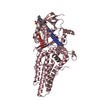
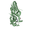
Mass: 41828.742 Da / Num. of mol.: 1 Fragment: Truncation of C-terminal polypro by V8-protease, UNP residues 2-373:VAL18ILE confirmed by MS Source method: isolated from a natural source Source: (natural) Todarodes pacificus (Japanese flying squid) References: UniProt: P31356
Mass: 256.424 Da / Num. of mol.: 2 / Source method: obtained synthetically / Formula: C16H32O2
Has protein modification
Y
Sequence details
THERE IS CONFLICTS BETWEEN SEQRES(ILE A 17) AND SEQUENCE DATABASE (VAL). THE AUTHORS CONFIRMED ...THERE IS CONFLICTS BETWEEN SEQRES(ILE A 17) AND SEQUENCE DATABASE (VAL). THE AUTHORS CONFIRMED EXPERIMENTALLY BY N-TERMINAL AMINO ACID SEQUENCING, AND THE SEQRES IS CORRECT AND IS THE TRUE IDENTITY OF THIS RESIDUE AND IS NATURAL VARIANT.
-
Experimental details
-
Experiment
Experiment
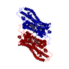
Method: X-RAY DIFFRACTION / Number of used crystals: 1
-
Sample preparation
Crystal
Density Matthews: 3.9 Å3/Da / Density % sol: 68.43 %
Crystal grow
Temperature: 293 K / Method: vapor diffusion, hanging drop / pH: 7 Details: 28% PEG400, 0.1M HEPES, 8% ethylene glycol, pH7.0, VAPOR DIFFUSION, HANGING DROP, temperature 293K
In the structure databanks used in Yorodumi, some data are registered as the other names, "COVID-19 virus" and "2019-nCoV". Here are the details of the virus and the list of structure data.
Jan 31, 2019. EMDB accession codes are about to change! (news from PDBe EMDB page)
EMDB accession codes are about to change! (news from PDBe EMDB page)
The allocation of 4 digits for EMDB accession codes will soon come to an end. Whilst these codes will remain in use, new EMDB accession codes will include an additional digit and will expand incrementally as the available range of codes is exhausted. The current 4-digit format prefixed with “EMD-” (i.e. EMD-XXXX) will advance to a 5-digit format (i.e. EMD-XXXXX), and so on. It is currently estimated that the 4-digit codes will be depleted around Spring 2019, at which point the 5-digit format will come into force.
The EM Navigator/Yorodumi systems omit the EMD- prefix.
Related info.:Q: What is EMD? / ID/Accession-code notation in Yorodumi/EM Navigator
Yorodumi is a browser for structure data from EMDB, PDB, SASBDB, etc.
This page is also the successor to EM Navigator detail page, and also detail information page/front-end page for Omokage search.
The word "yorodu" (or yorozu) is an old Japanese word meaning "ten thousand". "mi" (miru) is to see.
Related info.:EMDB / PDB / SASBDB / Comparison of 3 databanks / Yorodumi Search / Aug 31, 2016. New EM Navigator & Yorodumi / Yorodumi Papers / Jmol/JSmol / Function and homology information / Changes in new EM Navigator and Yorodumi
 Movie
Movie Controller


 Open data
 Basic information
 Components
 Keywords
 Function and homology information
 Todarodes pacificus (Japanese flying squid)
 X-RAY DIFFRACTION /
  Authors
 Citation
 Structure visualization
 Downloads & links
 Other downloads
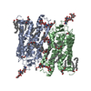


 PDBj
PDBj


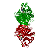
 Assembly

 Mass: 284.436 Da / Num. of mol.: 1 / Source method: obtained synthetically / Formula: C20H28O
 Mass: 256.424 Da / Num. of mol.: 2 / Source method: obtained synthetically / Formula: C16H32O2
 Sample preparation
 / Beamline: BL45XU / Wavelength: 0.9795 Å
 Processing