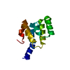
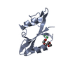
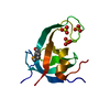
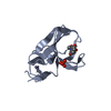
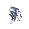
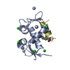
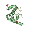
Entry Database : PDB / ID : 2x04Title Crystal structure of the PABC-TNRC6C complex POLYADENYLATE-BINDING PROTEIN 1 TRINUCLEOTIDE REPEAT-CONTAINING GENE 6C PROTEIN Keywords / / / / / / / / / / / / / Function / homology Function Domain/homology Component
/ / / / / / / / / / / / / / / / / / / / / / / / / / / / / / / / / / / / / / / / / / / / / / / / / / / / / / / / / / / / / / / / / / / / / / / / / / / / / / / / / / / / / / / / / / / / / / / / / / / / / / / / / / / / / / / / / / / Biological species HOMO SAPIENS (human)Method / / / Resolution : 1.49 Å Authors Jinek, M. / Fabian, M.R. / Coyle, S.M. / Sonenberg, N. / Doudna, J.A. Journal : Nat.Struct.Mol.Biol. / Year : 2010Title : Structural Insights Into the Human Gw182-Pabc Interaction in Microrna-Mediated DeadenylationAuthors : Jinek, M. / Fabian, M.R. / Coyle, S.M. / Sonenberg, N. / Doudna, J.A. History Deposition Dec 4, 2009 Deposition site / Processing site Revision 1.0 Jan 19, 2010 Provider / Type Revision 1.1 May 8, 2011 Group Revision 1.2 Jul 13, 2011 Group Revision 1.3 May 8, 2024 Group Data collection / Database references ... Data collection / Database references / Derived calculations / Other Category chem_comp_atom / chem_comp_bond ... chem_comp_atom / chem_comp_bond / database_2 / pdbx_database_status / struct_site Item _database_2.pdbx_DOI / _database_2.pdbx_database_accession ... _database_2.pdbx_DOI / _database_2.pdbx_database_accession / _pdbx_database_status.status_code_sf / _struct_site.pdbx_auth_asym_id / _struct_site.pdbx_auth_comp_id / _struct_site.pdbx_auth_seq_id
Show all Show less
 Movie
Movie Controller


 Open data
 Basic information
 Components
 Keywords
 Function and homology information
 HOMO SAPIENS (human)
 X-RAY DIFFRACTION /
  Authors
 Citation
 Structure visualization
 Downloads & links
 Other downloads
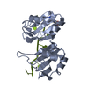


 PDBj
PDBj


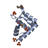
 Assembly


 Mass: 96.063 Da / Num. of mol.: 2 / Source method: obtained synthetically / Formula: SO4
 Mass: 18.015 Da / Num. of mol.: 217 / Source method: isolated from a natural source / Formula: H2O
 Sample preparation
 / Beamline: 8.2.1 / Wavelength: 0.999954
  Processing