 Movie
Movie Controller

[English] 日本語
 Yorodumi
- PDB-1jgn: Solution structure of the C-terminal PABC domain of human poly(A)... -
+ Open data

- Basic information
| Entry | Database: PDB / ID: 1jgn | ||||||
|---|---|---|---|---|---|---|---|
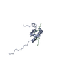
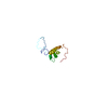
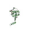
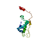
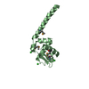
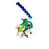
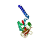
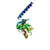
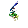
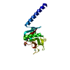
| Title | Solution structure of the C-terminal PABC domain of human poly(A)-binding protein in complex with the peptide from Paip2 | ||||||
 Components Components |
| ||||||
 Keywords Keywords | RNA BINDING PROTEIN / all-helical domain / protein-peptide complex | ||||||
| Function / homology |  Function and homology information Function and homology informationmCRD-mediated mRNA stability complex / negative regulation of nuclear-transcribed mRNA catabolic process, nonsense-mediated decay / negative regulation of nuclear-transcribed mRNA catabolic process, deadenylation-dependent decay / CRD-mediated mRNA stabilization / translation activator activity / Z-decay: degradation of maternal mRNAs by zygotically expressed factors / Deadenylation of mRNA / poly(A) binding / M-decay: degradation of maternal mRNAs by maternally stored factors / positive regulation of cytoplasmic translation ...mCRD-mediated mRNA stability complex / negative regulation of nuclear-transcribed mRNA catabolic process, nonsense-mediated decay / negative regulation of nuclear-transcribed mRNA catabolic process, deadenylation-dependent decay / CRD-mediated mRNA stabilization / translation activator activity / Z-decay: degradation of maternal mRNAs by zygotically expressed factors / Deadenylation of mRNA / poly(A) binding / M-decay: degradation of maternal mRNAs by maternally stored factors / positive regulation of cytoplasmic translation / regulation of long-term synaptic potentiation / regulatory ncRNA-mediated gene silencing / nuclear-transcribed mRNA catabolic process, nonsense-mediated decay / mRNA stabilization / positive regulation of nuclear-transcribed mRNA catabolic process, deadenylation-dependent decay / poly(U) RNA binding / positive regulation of nuclear-transcribed mRNA poly(A) tail shortening / Translation initiation complex formation / cell leading edge / Nonsense Mediated Decay (NMD) independent of the Exon Junction Complex (EJC) / L13a-mediated translational silencing of Ceruloplasmin expression / positive regulation of viral genome replication / Nonsense Mediated Decay (NMD) enhanced by the Exon Junction Complex (EJC) / catalytic step 2 spliceosome / translation repressor activity / negative regulation of translational initiation / mRNA regulatory element binding translation repressor activity / mRNA 3'-UTR binding / AUF1 (hnRNP D0) binds and destabilizes mRNA / mRNA splicing, via spliceosome / Regulation of expression of SLITs and ROBOs / cytoplasmic stress granule / memory / cytoplasmic ribonucleoprotein granule / lamellipodium / spermatogenesis / negative regulation of translation / translation / ribonucleoprotein complex / focal adhesion / mRNA binding / RNA binding / extracellular exosome / membrane / nucleus / cytoplasm / cytosol Similarity search - Function | ||||||
| Biological species |  Homo sapiens (human) Homo sapiens (human) | ||||||
| Method | SOLUTION NMR / simulated annealing | ||||||
 Authors Authors | Kozlov, G. / Siddiqui, N. / Coillet-Matillon, S. / Ekiel, I. / Gehring, K. | ||||||
 Citation Citation | Journal: EMBO J. / Year: 2004 Title: Structural basis of ligand recognition by PABC, a highly specific peptide-binding domain found in poly(A)-binding protein and a HECT ubiquitin ligase Authors: Kozlov, G. / De Crescenzo, G. / Lim, N.S. / Siddiqui, N. / Fantus, D. / Kahvejian, A. / Trempe, J.F. / Elias, D. / Ekiel, I. / Sonenberg, N. / O'Connor-McCourt, M. / Gehring, K. #1: Journal: Proc.Natl.Acad.Sci.USA / Year: 2001Title: Structure and function of the C-terminal PABC domain of human poly(A)-binding protein Authors: Kozlov, G. / Trempe, J.F. / Khaleghpour, K. / Kahvejian, A. / Ekiel, I. / Gehring, K. | ||||||
| History |
|
- Structure visualization
| Structure viewer | Molecule: MolmilJmol/JSmol |
|---|
- Downloads & links
-Download
| PDBx/mmCIF format | 1jgn.cif.gz | 1 MB | Display | PDBx/mmCIF format |
|---|---|---|---|---|
| PDB format | pdb1jgn.ent.gz | 880.6 KB | Display | PDB format |
| PDBx/mmJSON format | 1jgn.json.gz | Tree view | PDBx/mmJSON format | |
| Others |  Other downloads Other downloads |
-Validation report
| Arichive directory | https://data.pdbj.org/pub/pdb/validation_reports/jg/1jgnftp://data.pdbj.org/pub/pdb/validation_reports/jg/1jgn | HTTPS FTP |
|---|
-Related structure data
-Links
 PDBj
PDBj


- Assembly
| Deposited unit | 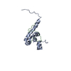
| |||||||||
|---|---|---|---|---|---|---|---|---|---|---|
| 1 |
| |||||||||
| NMR ensembles |
|
-Components
| #1: Protein | Mass: 10347.936 Da / Num. of mol.: 1 / Fragment: C-terminal domain Source method: isolated from a genetically manipulated source Source: (gene. exp.) Homo sapiens (human) / Gene: PABPC1 / Plasmid: pGEX-6P-1 / Production host:  |
|---|---|
| #2: Protein/peptide | Mass: 2390.755 Da / Num. of mol.: 1 / Fragment: C-terminal 22 residues Source method: isolated from a genetically manipulated source Source: (gene. exp.) Homo sapiens (human) / Plasmid: pGEX-6P-1 / Production host: |
-Experimental details
-Experiment
| Experiment | Method: SOLUTION NMR | ||||||||||||||||||||||||||||
|---|---|---|---|---|---|---|---|---|---|---|---|---|---|---|---|---|---|---|---|---|---|---|---|---|---|---|---|---|---|
| NMR experiment |
| ||||||||||||||||||||||||||||
| NMR details | Text: The structure was determined using standard triple-resonance and homonuclear techniques |
- Sample preparation
| Details |
| |||||||||||||||||||||
|---|---|---|---|---|---|---|---|---|---|---|---|---|---|---|---|---|---|---|---|---|---|---|
| Sample conditions | Ionic strength: 0.1M NaCl / pH: 6.3 / Pressure: ambient / Temperature: 303 K | |||||||||||||||||||||
| Crystal grow | *PLUS Method: other / Details: NMR |
-NMR measurement
| Radiation | Protocol: SINGLE WAVELENGTH / Monochromatic (M) / Laue (L): M | |||||||||||||||
|---|---|---|---|---|---|---|---|---|---|---|---|---|---|---|---|---|
| Radiation wavelength | Relative weight: 1 | |||||||||||||||
| NMR spectrometer |
|
- Processing
| NMR software |
| ||||||||||||||||||||||||
|---|---|---|---|---|---|---|---|---|---|---|---|---|---|---|---|---|---|---|---|---|---|---|---|---|---|
| Refinement | Method: simulated annealing / Software ordinal: 1 Details: The structures are based on 2058 non-redundant NOE-derived distance constraints, 106 dihedral angle restraints, and 35 hydrogen bonds. | ||||||||||||||||||||||||
| NMR representative | Selection criteria: lowest energy | ||||||||||||||||||||||||
| NMR ensemble | Conformer selection criteria: structures with the lowest energy Conformers calculated total number: 200 / Conformers submitted total number: 30 |
