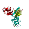
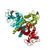
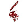
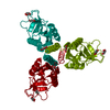
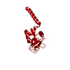
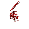
Signal regulatory protein family interactions / Surfactant metabolism / Toll Like Receptor 4 (TLR4) Cascade / Regulation of TLR by endogenous ligand / opsonization / response to interleukin-6 / respiratory gaseous exchange by respiratory system / response to epidermal growth factor / response to vitamin A / response to hyperoxia ...Signal regulatory protein family interactions / Surfactant metabolism / Toll Like Receptor 4 (TLR4) Cascade / Regulation of TLR by endogenous ligand / opsonization / response to interleukin-6 / respiratory gaseous exchange by respiratory system / response to epidermal growth factor / response to vitamin A / response to hyperoxia / response to retinoic acid / multivesicular body / cellular response to nitric oxide / response to hormone / positive regulation of phagocytosis / response to glucocorticoid / cellular response to mechanical stimulus / circadian rhythm / carbohydrate binding / response to lipopolysaccharide / response to hypoxia / : / metal ion binding Similarity search - Function
Collectin, C-type lectin-like domain / : / C-type lectin, conserved site / C-type lectin domain signature. / Mannose-Binding Protein A; Chain A / Mannose-Binding Protein A, subunit A / Lectin C-type domain / C-type lectin domain profile. / C-type lectin-like / C-type lectin (CTL) or carbohydrate-recognition domain (CRD) ...Collectin, C-type lectin-like domain / : / C-type lectin, conserved site / C-type lectin domain signature. / Mannose-Binding Protein A; Chain A / Mannose-Binding Protein A, subunit A / Lectin C-type domain / C-type lectin domain profile. / C-type lectin-like / C-type lectin (CTL) or carbohydrate-recognition domain (CRD) / C-type lectin-like/link domain superfamily / C-type lectin fold / Roll / Alpha Beta Similarity search - Domain/homology
Mass: 18.015 Da / Num. of mol.: 138 / Source method: isolated from a natural source / Formula: H2O
Has protein modification
Y
-
Experimental details
-
Experiment
Experiment
Method: X-RAY DIFFRACTION / Number of used crystals: 1
-
Sample preparation
Crystal
Density Matthews: 3.68 Å3/Da / Density % sol: 66.61 %
Crystal grow
Temperature: 290 K / Method: vapor diffusion, hanging drop / pH: 6 Details: Crystals were grown in hanging drops over reservoirs containing 10 mM sodium cacodylate (pH 6.0), 10 mM calcium acetate, and 10% (w/v) PEG 20,000
Protocol: SINGLE WAVELENGTH / Monochromatic (M) / Laue (L): M / Scattering type: x-ray
Radiation wavelength
Wavelength: 1.5418 Å / Relative weight: 1
Reflection
Resolution: 1.7→90 Å / Num. obs: 26014 / % possible obs: 96.7 % / Redundancy: 4.6 % / Biso Wilson estimate: 27.12 Å2 / Rmerge(I) obs: 0.027 / Χ2: 1.065 / Net I/av σ(I): 40.621 / Net I/σ(I): 45.2 / Num. measured all: 120336
Reflection shell
Diffraction-ID: 1 / Rejects: _
Resolution (Å)
Redundancy (%)
Rmerge(I) obs
Num. unique all
Χ2
% possible all
1.7-1.76
1.8
0.228
1963
1.463
74.1
1.76-1.83
2.5
0.181
2586
1.481
97.7
1.83-1.91
4.5
0.141
2692
1.21
100
1.91-2.02
5.2
0.1
2670
1.109
100
2.02-2.14
5.2
0.068
2693
1.152
100
2.14-2.31
5.2
0.05
2661
1.145
100
2.31-2.54
5.3
0.038
2693
1.105
99.9
2.54-2.91
5.3
0.03
2683
0.942
99.9
2.91-3.66
5.3
0.022
2700
0.718
99.5
3.662-90
5.1
0.02
2673
0.977
95.8
-
Processing
Software
Name
Version
Classification
DENZO
datareduction
SCALEPACK
datascaling
PHENIX
refinement
PDB_EXTRACT
3.15
dataextraction
Refinement
Resolution: 1.8→14.645 Å / FOM work R set: 0.897 / SU ML: 0.15 / Cross valid method: THROUGHOUT / σ(F): 0 / Phase error: 17.15 / Stereochemistry target values: ML
Rfactor
Num. reflection
% reflection
Rfree
0.1795
1707
7.65 %
Rwork
0.162
20614
-
obs
0.1635
22321
98.7 %
Solvent computation
Shrinkage radii: 0.9 Å / VDW probe radii: 1.11 Å / Solvent model: FLAT BULK SOLVENT MODEL
In the structure databanks used in Yorodumi, some data are registered as the other names, "COVID-19 virus" and "2019-nCoV". Here are the details of the virus and the list of structure data.
Jan 31, 2019. EMDB accession codes are about to change! (news from PDBe EMDB page)
EMDB accession codes are about to change! (news from PDBe EMDB page)
The allocation of 4 digits for EMDB accession codes will soon come to an end. Whilst these codes will remain in use, new EMDB accession codes will include an additional digit and will expand incrementally as the available range of codes is exhausted. The current 4-digit format prefixed with “EMD-” (i.e. EMD-XXXX) will advance to a 5-digit format (i.e. EMD-XXXXX), and so on. It is currently estimated that the 4-digit codes will be depleted around Spring 2019, at which point the 5-digit format will come into force.
The EM Navigator/Yorodumi systems omit the EMD- prefix.
Related info.:Q: What is EMD? / ID/Accession-code notation in Yorodumi/EM Navigator
Yorodumi is a browser for structure data from EMDB, PDB, SASBDB, etc.
This page is also the successor to EM Navigator detail page, and also detail information page/front-end page for Omokage search.
The word "yorodu" (or yorozu) is an old Japanese word meaning "ten thousand". "mi" (miru) is to see.
Related info.:EMDB / PDB / SASBDB / Comparison of 3 databanks / Yorodumi Search / Aug 31, 2016. New EM Navigator & Yorodumi / Yorodumi Papers / Jmol/JSmol / Function and homology information / Changes in new EM Navigator and Yorodumi
 Movie
Movie Controller


 Open data
 Basic information
 Components
 Keywords
 Function and homology information
 X-RAY DIFFRACTION / Resolution: 1.8 Å
  Authors
 United States, 1items
  Citation
 Structure visualization
 Downloads & links
 Other downloads


 PDBj
PDBj


 Assembly
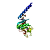
 Trichoplusia ni (cabbage looper) / References: UniProt: P08427
 Mass: 40.078 Da / Num. of mol.: 1 / Source method: obtained synthetically / Formula: Ca
 Mass: 18.015 Da / Num. of mol.: 138 / Source method: isolated from a natural source / Formula: H2O
 Sample preparation
 Processing