[English] 日本語
Yorodumi
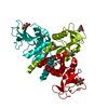
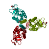
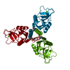
- PDB-1r13: Carbohydrate recognition and neck domains of surfactant protein A... -

+
Open data


ID or keywords:

Loading...

-
Basic information

Entry
Database: PDB / ID: 1r13
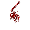
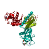
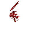
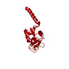
TitleCarbohydrate recognition and neck domains of surfactant protein A (SP-A)
ComponentsPulmonary surfactant-associated protein A
KeywordsSUGAR BINDING PROTEIN / CRD / alpha helical neck region
Function / homology
Function and homology information


Signal regulatory protein family interactions / Surfactant metabolism / Toll Like Receptor 4 (TLR4) Cascade / Regulation of TLR by endogenous ligand / opsonization / response to interleukin-6 / respiratory gaseous exchange by respiratory system / collagen trimer / response to epidermal growth factor / response to vitamin A ...Signal regulatory protein family interactions / Surfactant metabolism / Toll Like Receptor 4 (TLR4) Cascade / Regulation of TLR by endogenous ligand / opsonization / response to interleukin-6 / respiratory gaseous exchange by respiratory system / collagen trimer / response to epidermal growth factor / response to vitamin A / cellular response to nitric oxide / response to hyperoxia / response to retinoic acid / positive regulation of phagocytosis / response to glucocorticoid / multivesicular body / response to hormone / circadian rhythm / cellular response to mechanical stimulus / carbohydrate binding / response to lipopolysaccharide / response to hypoxia / extracellular space / metal ion binding
Similarity search - Function
Collectin, C-type lectin-like domain / C-type lectin, conserved site / C-type lectin domain signature. / Mannose-Binding Protein A; Chain A / Mannose-Binding Protein A, subunit A / Lectin C-type domain / C-type lectin domain profile. / C-type lectin-like / C-type lectin (CTL) or carbohydrate-recognition domain (CRD) / C-type lectin-like/link domain superfamily ...Collectin, C-type lectin-like domain / C-type lectin, conserved site / C-type lectin domain signature. / Mannose-Binding Protein A; Chain A / Mannose-Binding Protein A, subunit A / Lectin C-type domain / C-type lectin domain profile. / C-type lectin-like / C-type lectin (CTL) or carbohydrate-recognition domain (CRD) / C-type lectin-like/link domain superfamily / C-type lectin fold / Roll / Alpha Beta
Similarity search - Domain/homology
Pulmonary surfactant-associated protein A
Similarity search - Component
Biological speciesRattus norvegicus (Norway rat)
MethodX-RAY DIFFRACTION / SYNCHROTRON / MAD / Resolution: 2.1 Å
AuthorsHead, J.F. / Mealy, T.R. / McCormack, F.X. / Seaton, B.A.
CitationJournal: J.Biol.Chem. / Year: 2003
Title: Crystal structure of trimeric carbohydrate recognition and neck domains of surfactant protein A
Authors: Head, J.F. / Mealy, T.R. / McCormack, F.X. / Seaton, B.A.
History
DepositionSep 23, 2003Deposition site: RCSB / Processing site: RCSB
Revision 1.0Nov 18, 2003Provider: repository / Type: Initial release
Revision 1.1Apr 29, 2008Group: Version format compliance
Revision 1.2Jul 13, 2011Group: Derived calculations / Version format compliance
Revision 1.3Oct 27, 2021Group: Database references / Derived calculations
Category: database_2 / pdbx_struct_conn_angle ...database_2 / pdbx_struct_conn_angle / struct_conn / struct_ref_seq_dif / struct_site
Item: _database_2.pdbx_DOI / _database_2.pdbx_database_accession ..._database_2.pdbx_DOI / _database_2.pdbx_database_accession / _pdbx_struct_conn_angle.ptnr1_auth_comp_id / _pdbx_struct_conn_angle.ptnr1_auth_seq_id / _pdbx_struct_conn_angle.ptnr1_label_asym_id / _pdbx_struct_conn_angle.ptnr1_label_atom_id / _pdbx_struct_conn_angle.ptnr1_label_comp_id / _pdbx_struct_conn_angle.ptnr1_label_seq_id / _pdbx_struct_conn_angle.ptnr3_auth_comp_id / _pdbx_struct_conn_angle.ptnr3_auth_seq_id / _pdbx_struct_conn_angle.ptnr3_label_asym_id / _pdbx_struct_conn_angle.ptnr3_label_atom_id / _pdbx_struct_conn_angle.ptnr3_label_comp_id / _pdbx_struct_conn_angle.ptnr3_label_seq_id / _pdbx_struct_conn_angle.value / _struct_conn.pdbx_dist_value / _struct_conn.ptnr1_auth_comp_id / _struct_conn.ptnr1_auth_seq_id / _struct_conn.ptnr1_label_asym_id / _struct_conn.ptnr1_label_atom_id / _struct_conn.ptnr1_label_comp_id / _struct_conn.ptnr1_label_seq_id / _struct_conn.ptnr2_auth_comp_id / _struct_conn.ptnr2_auth_seq_id / _struct_conn.ptnr2_label_asym_id / _struct_conn.ptnr2_label_atom_id / _struct_conn.ptnr2_label_comp_id / _struct_conn.ptnr2_label_seq_id / _struct_ref_seq_dif.details / _struct_site.pdbx_auth_asym_id / _struct_site.pdbx_auth_comp_id / _struct_site.pdbx_auth_seq_id

-
Structure visualization

Structure viewerMolecule:
MolmilJmol/JSmol

Downloads & links

-
Assembly

Deposited unit
A: Pulmonary surfactant-associated protein A
hetero molecules


Theoretical massNumber of molelcules
Total (without water)17,0044
Polymers16,6731
Non-polymers3313
Water2,108117
1
A: Pulmonary surfactant-associated protein A
hetero molecules

A: Pulmonary surfactant-associated protein A
hetero molecules

A: Pulmonary surfactant-associated protein A
hetero molecules


Theoretical massNumber of molelcules
Total (without water)51,01212
Polymers50,0183
Non-polymers9949
Water543
TypeNameSymmetry operationNumber
identity operation1_555x,y,z1
crystal symmetry operation2_655-y+1,x-y,z1
crystal symmetry operation3_665-x+y+1,-x+1,z1
Buried area5730 Å2
ΔGint-113 kcal/mol
Surface area21430 Å2
MethodPISA, PQS
Unit cell
Length a, b, c (Å)97.68, 97.68, 44.54
Angle α, β, γ (deg.)90.0, 90.0, 120.0
Int Tables number173
Space group name H-MP63

-
Components

#1: Protein Pulmonary surfactant-associated protein A / SP-A / PSP-A / PSAP


Mass: 16672.580 Da / Num. of mol.: 1 / Fragment: CRD and neck domains / Mutation: N187S
Source method: isolated from a genetically manipulated source
Source: (gene. exp.) Rattus norvegicus (Norway rat) / Gene: SFTPA1 OR SFTPA OR SFTP1 OR SFTP-1 / Plasmid: PVL 1392 / Genus (production host): Trichoplusia / Production host: 
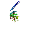
Trichoplusia (butterflies/moths) / References: UniProt: P08427
#2: Chemical ChemComp-CA / CALCIUM ION


Mass: 40.078 Da / Num. of mol.: 1 / Source method: obtained synthetically / Formula: Ca
#3: Chemical ChemComp-SO4 / SULFATE ION


Mass: 96.063 Da / Num. of mol.: 1 / Source method: obtained synthetically / Formula: SO4
#4: Chemical ChemComp-MES / 2-(N-MORPHOLINO)-ETHANESULFONIC ACID


Mass: 195.237 Da / Num. of mol.: 1 / Source method: obtained synthetically / Formula: C6H13NO4S / Comment: pH buffer*YM
#5: Water ChemComp-HOH / water


Mass: 18.015 Da / Num. of mol.: 117 / Source method: isolated from a natural source / Formula: H2O

-
Experimental details

-
Experiment

ExperimentMethod: X-RAY DIFFRACTION / Number of used crystals: 1

-
Sample preparation

CrystalDensity Matthews: 3.68 Å3/Da / Density % sol: 66.56 %
Crystal growTemperature: 290 K / Method: vapor diffusion, sitting drop / pH: 6.5
Details: LiSO4, CaCl2, glycerol, trimannose, MES, pH 6.5, VAPOR DIFFUSION, SITTING DROP, temperature 290K
Crystal grow
*PLUS
Temperature: 17 ℃ / Method: vapor diffusion, sitting drop
Components of the solutions
*PLUS
IDConc.Common nameCrystal-IDSol-IDChemical formulaDetails
15 mMTris1drop
250 mM1dropNaCl
33 mg/mlprotein1drop
41.6 M1reservoirLiSO4
510 mM1reservoirCaCl2
67 %(w/v)glycerol1reservoir
7100 mMMES1reservoirpH6.5

-
Data collection

DiffractionMean temperature: 100 K
Diffraction sourceSource: SYNCHROTRON / Site: NSLS / Beamline: X8C / Wavelength: 1.072 Å
DetectorType: ADSC QUANTUM 4 / Detector: CCD / Date: Oct 8, 2000
RadiationProtocol: MAD / Monochromatic (M) / Laue (L): M / Scattering type: x-ray
Radiation wavelengthWavelength: 1.072 Å / Relative weight: 1
ReflectionResolution: 2.1→50 Å / Num. all: 14375 / Num. obs: 14373 / % possible obs: 99.5 % / Observed criterion σ(F): -1 / Observed criterion σ(I): -1 / Redundancy: 13.1 % / Rmerge(I) obs: 0.059 / Net I/σ(I): 25.4
Reflection shellResolution: 2.1→2.18 Å / Rmerge(I) obs: 0.299 / Mean I/σ(I) obs: 6.6 / Num. unique all: 1382 / % possible all: 98.3
Reflection
*PLUS
Num. measured all: 189251
Reflection shell
*PLUS
% possible obs: 98.3 %

-
Processing

Software
NameVersionClassification
DENZOdata reduction
SCALEPACKdata scaling
SOLVEphasing
CNS1refinement
RefinementMethod to determine structure: MAD / Resolution: 2.1→50 Å / Isotropic thermal model: isotropic / Cross valid method: THROUGHOUT / σ(F): 0 / σ(I): 0 / Stereochemistry target values: Engh & Huber
RfactorNum. reflection% reflectionSelection details
Rfree0.249 1421 -random
Rwork0.221 ---
all0.221 14360 --
obs0.221 14049 97.8 %-
Refinement stepCycle: LAST / Resolution: 2.1→50 Å
ProteinNucleic acidLigandSolventTotal
Num. atoms1143 0 18 117 1278
Refine LS restraints
*PLUS
Refine-IDTypeDev ideal
X-RAY DIFFRACTIONbond_d0.00625
X-RAY DIFFRACTIONangle_d
X-RAY DIFFRACTIONangle_deg1.212

+
About Yorodumi

-
News

-
Feb 9, 2022. New format data for meta-information of EMDB entries

New format data for meta-information of EMDB entries

  • Version 3 of the EMDB header file is now the official format.
  • The previous official version 1.9 will be removed from the archive.

Related info.:EMDB header

External links:wwPDB to switch to version 3 of the EMDB data model

-
Aug 12, 2020. Covid-19 info

Covid-19 info

URL: https://pdbjlvh1.pdbj.org/emnavi/covid19.php

New page: Covid-19 featured information page in EM Navigator.

Related info.:Covid-19 info / Mar 5, 2020. Novel coronavirus structure data

+
Mar 5, 2020. Novel coronavirus structure data

Novel coronavirus structure data

Related info.:Yorodumi Speices / Aug 12, 2020. Covid-19 info

External links:COVID-19 featured content - PDBj / Molecule of the Month (242):Coronavirus Proteases

+
Jan 31, 2019. EMDB accession codes are about to change! (news from PDBe EMDB page)

EMDB accession codes are about to change! (news from PDBe EMDB page)

  • The allocation of 4 digits for EMDB accession codes will soon come to an end. Whilst these codes will remain in use, new EMDB accession codes will include an additional digit and will expand incrementally as the available range of codes is exhausted. The current 4-digit format prefixed with “EMD-” (i.e. EMD-XXXX) will advance to a 5-digit format (i.e. EMD-XXXXX), and so on. It is currently estimated that the 4-digit codes will be depleted around Spring 2019, at which point the 5-digit format will come into force.
  • The EM Navigator/Yorodumi systems omit the EMD- prefix.

Related info.:Q: What is EMD? / ID/Accession-code notation in Yorodumi/EM Navigator

External links:EMDB Accession Codes are Changing Soon! / Contact to PDBj

+
Jul 12, 2017. Major update of PDB

Major update of PDB

  • wwPDB released updated PDB data conforming to the new PDBx/mmCIF dictionary.
  • This is a major update changing the version number from 4 to 5, and with Remediation, in which all the entries are updated.
  • In this update, many items about electron microscopy experimental information are reorganized (e.g. em_software).
  • Now, EM Navigator and Yorodumi are based on the updated data.

External links:wwPDB Remediation / Enriched Model Files Conforming to OneDep Data Standards Now Available in the PDB FTP Archive

-
Yorodumi

Thousand views of thousand structures

  • Yorodumi is a browser for structure data from EMDB, PDB, SASBDB, etc.
  • This page is also the successor to EM Navigator detail page, and also detail information page/front-end page for Omokage search.
  • The word "yorodu" (or yorozu) is an old Japanese word meaning "ten thousand". "mi" (miru) is to see.

Related info.:EMDB / PDB / SASBDB / Comparison of 3 databanks / Yorodumi Search / Aug 31, 2016. New EM Navigator & Yorodumi / Yorodumi Papers / Jmol/JSmol / Function and homology information / Changes in new EM Navigator and Yorodumi

Read more