[English] 日本語
Yorodumi
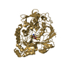
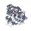
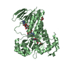
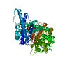
- PDB-2wmg: Crystal structure of the catalytic module of a family 98 glycosid... -

+
Open data


ID or keywords:

Loading...

-
Basic information

Entry
Database: PDB / ID: 2wmg
Title
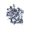
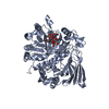
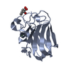
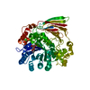
Crystal structure of the catalytic module of a family 98 glycoside hydrolase from Streptococcus pneumoniae TIGR4 (Sp4GH98) in complex with the LewisY pentasaccharide blood group antigen.
ComponentsFUCOLECTIN-RELATED PROTEIN
KeywordsHYDROLASE / GLYCOSIDE HYDROLASE / STREPTOCOCCUS PNEUMONIAE / LEWISY BLOOD GROUP ANTIGEN / FUCOSE UTILIZATION
Function / homology
Function and homology information


regulation of complement activation, lectin pathway / regulation of cellular defense response / fucose binding / carbohydrate metabolic process / metal ion binding
Similarity search - Function
family 98 glycoside hydrolase / Fucolectin tachylectin-4 pentraxin-1 / eel-Fucolectin Tachylectin-4 Pentaxrin-1 Domain / Glycosyl hydrolase family 98, C-terminal / Glycosyl hydrolase family 98, central domain / Glycosyl hydrolase family 98 / Glycosyl hydrolase family 98 C-terminal domain / arginine biosynthesis bifunctional protein fold / Polysaccharide lyase family 8-like, C-terminal / Chondroitinase Ac; Chain A, domain 3 ...family 98 glycoside hydrolase / Fucolectin tachylectin-4 pentraxin-1 / eel-Fucolectin Tachylectin-4 Pentaxrin-1 Domain / Glycosyl hydrolase family 98, C-terminal / Glycosyl hydrolase family 98, central domain / Glycosyl hydrolase family 98 / Glycosyl hydrolase family 98 C-terminal domain / arginine biosynthesis bifunctional protein fold / Polysaccharide lyase family 8-like, C-terminal / Chondroitinase Ac; Chain A, domain 3 / Polysaccharide lyase family 8-like, C-terminal / Coagulation factors 5/8 type C domain (FA58C) profile. / F5/8 type C domain / Coagulation factor 5/8 C-terminal domain / Galactose-binding-like domain superfamily / Glycosidases / TIM Barrel / Alpha-Beta Barrel / Sandwich / 2-Layer Sandwich / Mainly Beta / Alpha Beta
Similarity search - Domain/homology
Lewis Y antigen, beta anomer / Fucolectin-related protein / Fucolectin-related protein
Similarity search - Component
Biological speciesSTREPTOCOCCUS PNEUMONIAE (bacteria)
MethodX-RAY DIFFRACTION / SYNCHROTRON / OTHER / Resolution: 2.3 Å
AuthorsHiggins, M.A. / Whitworth, G.E. / El Warry, N. / Randriantsoa, M. / Samain, E. / Burke, R.D. / Vocadlo, D.J. / Boraston, A.B.
CitationJournal: J.Biol.Chem. / Year: 2009
Title: Differential Recognition and Hydrolysis of Host Carbohydrate-Antigens by Streptococcus Pneumoniae Family 98 Glycoside Hydrolases.
Authors: Higgins, M.A. / Whitworth, G.E. / El Warry, N. / Randriantsoa, M. / Samain, E. / Burke, R.D. / Vocadlo, D.J. / Boraston, A.B.
History
DepositionJun 30, 2009Deposition site: PDBE / Processing site: PDBE
Revision 1.0Jul 14, 2009Provider: repository / Type: Initial release
Revision 1.1May 8, 2011Group: Version format compliance
Revision 1.2Jul 13, 2011Group: Version format compliance
Revision 2.0Jul 29, 2020Group: Atomic model / Data collection ...Atomic model / Data collection / Derived calculations / Other / Structure summary
Category: atom_site / atom_site_anisotrop ...atom_site / atom_site_anisotrop / chem_comp / entity / entity_name_com / pdbx_branch_scheme / pdbx_chem_comp_identifier / pdbx_database_status / pdbx_entity_branch / pdbx_entity_branch_descriptor / pdbx_entity_branch_link / pdbx_entity_branch_list / pdbx_entity_nonpoly / pdbx_molecule_features / pdbx_nonpoly_scheme / pdbx_struct_assembly_gen / struct_asym / struct_conn / struct_site / struct_site_gen
Item: _atom_site.B_iso_or_equiv / _atom_site.Cartn_x ..._atom_site.B_iso_or_equiv / _atom_site.Cartn_x / _atom_site.Cartn_y / _atom_site.Cartn_z / _atom_site.auth_asym_id / _atom_site.auth_atom_id / _atom_site.auth_comp_id / _atom_site.auth_seq_id / _atom_site.label_asym_id / _atom_site.label_atom_id / _atom_site.label_comp_id / _atom_site.label_entity_id / _atom_site.type_symbol / _atom_site_anisotrop.U[1][1] / _atom_site_anisotrop.U[1][2] / _atom_site_anisotrop.U[1][3] / _atom_site_anisotrop.U[2][2] / _atom_site_anisotrop.U[2][3] / _atom_site_anisotrop.U[3][3] / _atom_site_anisotrop.pdbx_auth_asym_id / _atom_site_anisotrop.pdbx_auth_atom_id / _atom_site_anisotrop.pdbx_auth_comp_id / _atom_site_anisotrop.pdbx_auth_seq_id / _atom_site_anisotrop.pdbx_label_asym_id / _atom_site_anisotrop.pdbx_label_atom_id / _atom_site_anisotrop.pdbx_label_comp_id / _atom_site_anisotrop.type_symbol / _chem_comp.name / _chem_comp.type / _pdbx_database_status.status_code_sf / _pdbx_struct_assembly_gen.asym_id_list / _struct_conn.pdbx_dist_value / _struct_conn.pdbx_leaving_atom_flag / _struct_conn.ptnr1_auth_asym_id / _struct_conn.ptnr1_auth_comp_id / _struct_conn.ptnr1_auth_seq_id / _struct_conn.ptnr1_label_asym_id / _struct_conn.ptnr1_label_atom_id / _struct_conn.ptnr1_label_comp_id / _struct_conn.ptnr2_auth_asym_id / _struct_conn.ptnr2_auth_comp_id / _struct_conn.ptnr2_auth_seq_id / _struct_conn.ptnr2_label_asym_id / _struct_conn.ptnr2_label_atom_id / _struct_conn.ptnr2_label_comp_id
Description: Carbohydrate remediation / Provider: repository / Type: Remediation
Remark 700 SHEET DETERMINATION METHOD: DSSP THE SHEETS PRESENTED AS "AA" IN EACH CHAIN ON SHEET RECORDS BELOW ... SHEET DETERMINATION METHOD: DSSP THE SHEETS PRESENTED AS "AA" IN EACH CHAIN ON SHEET RECORDS BELOW IS ACTUALLY AN 9-STRANDED BARREL THIS IS REPRESENTED BY A 10-STRANDED SHEET IN WHICH THE FIRST AND LAST STRANDS ARE IDENTICAL.

-
Structure visualization

Structure viewerMolecule:
MolmilJmol/JSmol

Downloads & links

-
Assembly

Deposited unit
A: FUCOLECTIN-RELATED PROTEIN
hetero molecules


Theoretical massNumber of molelcules
Total (without water)67,4452
Polymers66,7701
Non-polymers6761
Water6,017334
1


  • Idetical with deposited unit
  • defined by author&software
TypeNameSymmetry operationNumber
identity operation1_555x,y,z1
MethodPQS
Unit cell
Length a, b, c (Å)50.860, 90.870, 116.230
Angle α, β, γ (deg.)90.00, 90.00, 90.00
Int Tables number19
Space group name H-MP212121

-
Components

#1: Protein FUCOLECTIN-RELATED PROTEIN / GLYCOSIDE HYDROLASE


Mass: 66769.758 Da / Num. of mol.: 1 / Fragment: CATALYTIC MODULE, RESIDUES 31-589 / Mutation: YES
Source method: isolated from a genetically manipulated source
Source: (gene. exp.) STREPTOCOCCUS PNEUMONIAE (bacteria) / Strain: TIGR4 / Plasmid: PET28A / Production host: ESCHERICHIA COLI (E. coli) / Strain (production host): BL21 STAR (DE3) / References: UniProt: Q97N96, UniProt: A0A0H2US34*PLUS
#2: Polysaccharide 
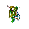
alpha-L-fucopyranose-(1-2)-beta-D-galactopyranose-(1-4)-[alpha-L-fucopyranose-(1-3)]2-acetamido-2- ...alpha-L-fucopyranose-(1-2)-beta-D-galactopyranose-(1-4)-[alpha-L-fucopyranose-(1-3)]2-acetamido-2-deoxy-beta-D-glucopyranose / Lewis Y antigen / beta anomer


Type: oligosaccharide, Oligosaccharide / Class: Antigen / Mass: 675.630 Da / Num. of mol.: 1
Source method: isolated from a genetically manipulated source
Details: oligosaccharide with branches / References: Lewis Y antigen, beta anomer
DescriptorTypeProgram
LFucpa1-2DGalpb1-4[LFucpa1-3]DGlcpNAcb1-ROHGlycam Condensed SequenceGMML 1.0
WURCS=2.0/3,4,3/[a2122h-1b_1-5_2*NCC/3=O][a1221m-1a_1-5][a2112h-1b_1-5]/1-2-3-2/a3-b1_a4-c1_c2-d1WURCSPDB2Glycan 1.1.0
[][b-D-GlcpNAc]{[(3+1)][a-L-Fucp]{}[(4+1)][b-D-Galp]{[(2+1)][a-L-Fucp]{}}}LINUCSPDB-CARE
#3: Water ChemComp-HOH / water / Water


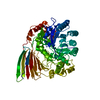
Mass: 18.015 Da / Num. of mol.: 334 / Source method: isolated from a natural source / Formula: H2O
Compound detailsENGINEERED RESIDUE IN CHAIN A, GLU 158 TO ALA

-
Experimental details

-
Experiment

ExperimentMethod: X-RAY DIFFRACTION

-
Sample preparation

CrystalDensity Matthews: 1.97 Å3/Da / Density % sol: 36.96 % / Description: NONE

-
Data collection

DiffractionMean temperature: 292 K
Diffraction sourceSource: SYNCHROTRON / Site: SSRL / Beamline: BL7-1 / Wavelength: 0.9214
DetectorType: ADSC QUANTUM 315r / Detector: CCD
RadiationProtocol: SINGLE WAVELENGTH / Monochromatic (M) / Laue (L): M / Scattering type: x-ray
Radiation wavelengthWavelength: 0.9214 Å / Relative weight: 1
ReflectionResolution: 2.28→40 Å / Num. obs: 22735 / % possible obs: 97.7 % / Observed criterion σ(I): 2 / Redundancy: 4.8 % / Rmerge(I) obs: 0.14 / Net I/σ(I): 11

-
Processing

SoftwareName: REFMAC / Version: 5.2.0019 / Classification: refinement
RefinementMethod to determine structure: OTHER
Starting model: NONE

Resolution: 2.3→30.29 Å / Cor.coef. Fo:Fc: 0.95 / Cor.coef. Fo:Fc free: 0.889 / SU B: 6.299 / SU ML: 0.156 / Cross valid method: THROUGHOUT / ESU R: 0.472 / ESU R Free: 0.249 / Stereochemistry target values: MAXIMUM LIKELIHOOD / Details: HYDROGENS HAVE BEEN ADDED IN THE RIDING POSITIONS.
RfactorNum. reflection% reflectionSelection details
Rfree0.22745 1226 5.1 %RANDOM
Rwork0.1564 ---
obs0.15998 22735 97.07 %-
Solvent computationIon probe radii: 0.8 Å / Shrinkage radii: 0.8 Å / VDW probe radii: 1.4 Å / Solvent model: MASK
Displacement parametersBiso mean: 12.945 Å2
Baniso -1Baniso -2Baniso -3
1-0.01 Å20 Å20 Å2
2--0 Å20 Å2
3----0.01 Å2
Refinement stepCycle: LAST / Resolution: 2.3→30.29 Å
ProteinNucleic acidLigandSolventTotal
Num. atoms4498 0 46 334 4878
Refine LS restraints
Refine-IDTypeDev idealDev ideal targetNumber
X-RAY DIFFRACTIONr_bond_refined_d0.010.0224679
X-RAY DIFFRACTIONr_bond_other_d
X-RAY DIFFRACTIONr_angle_refined_deg1.3041.9376378
X-RAY DIFFRACTIONr_angle_other_deg
X-RAY DIFFRACTIONr_dihedral_angle_1_deg6.5015551
X-RAY DIFFRACTIONr_dihedral_angle_2_deg36.4924.912228
X-RAY DIFFRACTIONr_dihedral_angle_3_deg14.19715762
X-RAY DIFFRACTIONr_dihedral_angle_4_deg18.0831515
X-RAY DIFFRACTIONr_chiral_restr0.090.2685
X-RAY DIFFRACTIONr_gen_planes_refined0.0040.023579
X-RAY DIFFRACTIONr_gen_planes_other
X-RAY DIFFRACTIONr_nbd_refined0.1920.22250
X-RAY DIFFRACTIONr_nbd_other
X-RAY DIFFRACTIONr_nbtor_refined0.3060.23124
X-RAY DIFFRACTIONr_nbtor_other
X-RAY DIFFRACTIONr_xyhbond_nbd_refined0.1260.2329
X-RAY DIFFRACTIONr_xyhbond_nbd_other
X-RAY DIFFRACTIONr_metal_ion_refined
X-RAY DIFFRACTIONr_metal_ion_other
X-RAY DIFFRACTIONr_symmetry_vdw_refined0.1780.253
X-RAY DIFFRACTIONr_symmetry_vdw_other
X-RAY DIFFRACTIONr_symmetry_hbond_refined0.2960.217
X-RAY DIFFRACTIONr_symmetry_hbond_other
X-RAY DIFFRACTIONr_symmetry_metal_ion_refined
X-RAY DIFFRACTIONr_symmetry_metal_ion_other
X-RAY DIFFRACTIONr_mcbond_it0.5151.52833
X-RAY DIFFRACTIONr_mcbond_other
X-RAY DIFFRACTIONr_mcangle_it0.86324483
X-RAY DIFFRACTIONr_mcangle_other
X-RAY DIFFRACTIONr_scbond_it1.35832175
X-RAY DIFFRACTIONr_scbond_other
X-RAY DIFFRACTIONr_scangle_it2.1124.51895
X-RAY DIFFRACTIONr_scangle_other
X-RAY DIFFRACTIONr_long_range_B_refined
X-RAY DIFFRACTIONr_long_range_B_other
X-RAY DIFFRACTIONr_rigid_bond_restr
X-RAY DIFFRACTIONr_sphericity_free
X-RAY DIFFRACTIONr_sphericity_bonded
LS refinement shellResolution: 2.3→2.359 Å / Total num. of bins used: 20
RfactorNum. reflection% reflection
Rfree0.281 89 -
Rwork0.172 1631 -
obs--97.07 %

+
About Yorodumi

-
News

-
Feb 9, 2022. New format data for meta-information of EMDB entries

New format data for meta-information of EMDB entries

  • Version 3 of the EMDB header file is now the official format.
  • The previous official version 1.9 will be removed from the archive.

Related info.:EMDB header

External links:wwPDB to switch to version 3 of the EMDB data model

-
Aug 12, 2020. Covid-19 info

Covid-19 info

URL: https://pdbj.org/emnavi/covid19.php

New page: Covid-19 featured information page in EM Navigator.

Related info.:Covid-19 info / Mar 5, 2020. Novel coronavirus structure data

+
Mar 5, 2020. Novel coronavirus structure data

Novel coronavirus structure data

Related info.:Yorodumi Speices / Aug 12, 2020. Covid-19 info

External links:COVID-19 featured content - PDBj / Molecule of the Month (242):Coronavirus Proteases

+
Jan 31, 2019. EMDB accession codes are about to change! (news from PDBe EMDB page)

EMDB accession codes are about to change! (news from PDBe EMDB page)

  • The allocation of 4 digits for EMDB accession codes will soon come to an end. Whilst these codes will remain in use, new EMDB accession codes will include an additional digit and will expand incrementally as the available range of codes is exhausted. The current 4-digit format prefixed with “EMD-” (i.e. EMD-XXXX) will advance to a 5-digit format (i.e. EMD-XXXXX), and so on. It is currently estimated that the 4-digit codes will be depleted around Spring 2019, at which point the 5-digit format will come into force.
  • The EM Navigator/Yorodumi systems omit the EMD- prefix.

Related info.:Q: What is EMD? / ID/Accession-code notation in Yorodumi/EM Navigator

External links:EMDB Accession Codes are Changing Soon! / Contact to PDBj

+
Jul 12, 2017. Major update of PDB

Major update of PDB

  • wwPDB released updated PDB data conforming to the new PDBx/mmCIF dictionary.
  • This is a major update changing the version number from 4 to 5, and with Remediation, in which all the entries are updated.
  • In this update, many items about electron microscopy experimental information are reorganized (e.g. em_software).
  • Now, EM Navigator and Yorodumi are based on the updated data.

External links:wwPDB Remediation / Enriched Model Files Conforming to OneDep Data Standards Now Available in the PDB FTP Archive

-
Yorodumi

Thousand views of thousand structures

  • Yorodumi is a browser for structure data from EMDB, PDB, SASBDB, etc.
  • This page is also the successor to EM Navigator detail page, and also detail information page/front-end page for Omokage search.
  • The word "yorodu" (or yorozu) is an old Japanese word meaning "ten thousand". "mi" (miru) is to see.

Related info.:EMDB / PDB / SASBDB / Comparison of 3 databanks / Yorodumi Search / Aug 31, 2016. New EM Navigator & Yorodumi / Yorodumi Papers / Jmol/JSmol / Function and homology information / Changes in new EM Navigator and Yorodumi

Read more