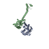
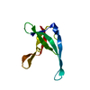
regulation of type II interferon-mediated signaling pathway / HSP90-CDC37 chaperone complex / positive regulation of type 2 mitophagy / regulation of cyclin-dependent protein serine/threonine kinase activity / protein kinase regulator activity / post-transcriptional regulation of gene expression / Drug-mediated inhibition of ERBB2 signaling / Resistance of ERBB2 KD mutants to trastuzumab / Resistance of ERBB2 KD mutants to sapitinib / Resistance of ERBB2 KD mutants to tesevatinib ...regulation of type II interferon-mediated signaling pathway / HSP90-CDC37 chaperone complex / positive regulation of type 2 mitophagy / regulation of cyclin-dependent protein serine/threonine kinase activity / protein kinase regulator activity / post-transcriptional regulation of gene expression / Drug-mediated inhibition of ERBB2 signaling / Resistance of ERBB2 KD mutants to trastuzumab / Resistance of ERBB2 KD mutants to sapitinib / Resistance of ERBB2 KD mutants to tesevatinib / Resistance of ERBB2 KD mutants to neratinib / Resistance of ERBB2 KD mutants to osimertinib / Resistance of ERBB2 KD mutants to afatinib / Resistance of ERBB2 KD mutants to AEE788 / Resistance of ERBB2 KD mutants to lapatinib / Drug resistance in ERBB2 TMD/JMD mutants / regulation of type I interferon-mediated signaling pathway / protein folding chaperone complex / RHOBTB2 GTPase cycle / protein targeting / heat shock protein binding / Signaling by ERBB2 / Constitutive Signaling by Overexpressed ERBB2 / Signaling by ERBB2 TMD/JMD mutants / Hsp90 protein binding / Constitutive Signaling by EGFRvIII / Signaling by ERBB2 ECD mutants / Signaling by ERBB2 KD Mutants / Regulation of necroptotic cell death / kinase binding / Downregulation of ERBB2 signaling / unfolded protein binding / Constitutive Signaling by Ligand-Responsive EGFR Cancer Variants / protein-folding chaperone binding / protein folding / scaffold protein binding / protein stabilization / protein kinase binding / extracellular exosome / cytoplasm / cytosol Similarity search - Function
Resolution: 1.88→42.14 Å / Cor.coef. Fo:Fc: 0.943 / Cor.coef. Fo:Fc free: 0.926 / SU B: 3.364 / SU ML: 0.104 / Cross valid method: THROUGHOUT / ESU R: 0.178 / ESU R Free: 0.156 / Stereochemistry target values: MAXIMUM LIKELIHOOD / Details: HYDROGENS HAVE BEEN ADDED IN THE RIDING POSITIONS.
Rfactor
Num. reflection
% reflection
Selection details
Rfree
0.246
536
4.9 %
RANDOM
Rwork
0.212
-
-
-
obs
0.213
10483
96.9 %
-
Solvent computation
Ion probe radii: 0.8 Å / Shrinkage radii: 0.8 Å / VDW probe radii: 1.2 Å / Solvent model: MASK
 Movie
Movie Controller


 Open data
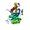
 Basic information
 Components
 Keywords
 Function and homology information
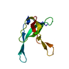
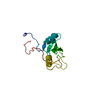
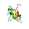
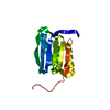
 HOMO SAPIENS (human)
 X-RAY DIFFRACTION /
  Authors
 Citation
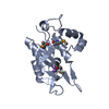
 Structure visualization
 Downloads & links
 Other downloads
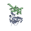
 PDBj
PDBj


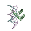
 Assembly

 Mass: 18.015 Da / Num. of mol.: 74 / Source method: isolated from a natural source / Formula: H2O
 Sample preparation
 Processing