 Movie
Movie Controller

[English] 日本語
 Yorodumi
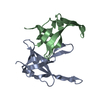
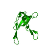
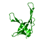
- PDB-1yhb: CRYSTAL STRUCTURES OF Y41H AND Y41F MUTANTS OF GENE V PROTEIN FRO... -
+ Open data

- Basic information
| Entry | Database: PDB / ID: 1yhb | ||||||
|---|---|---|---|---|---|---|---|
| Title | CRYSTAL STRUCTURES OF Y41H AND Y41F MUTANTS OF GENE V PROTEIN FROM FF PHAGE SUGGEST POSSIBLE PROTEIN-PROTEIN INTERACTIONS IN GVP-SSDNA COMPLEX | ||||||
 Components Components | GENE V PROTEIN | ||||||
 Keywords Keywords | DNA BINDING PROTEIN / DNA-BINDING PROTEIN | ||||||
| Function / homology |  Function and homology information Function and homology informationrolling circle single-stranded viral DNA replication / single-stranded DNA binding / DNA replication Similarity search - Function | ||||||
| Biological species |  Enterobacteria phage f1 (virus) Enterobacteria phage f1 (virus) | ||||||
| Method |  X-RAY DIFFRACTION / Resolution: 2.2 Å X-RAY DIFFRACTION / Resolution: 2.2 Å | ||||||
 Authors Authors | Guan, Y. / Zhang, H. / Konings, R.N.H. / Hilbers, C.W. / Terwilliger, T.C. / Wang, A.H.-J. | ||||||
 Citation Citation | Journal: Biochemistry / Year: 1994 Title: Crystal structures of Y41H and Y41F mutants of gene V protein from Ff phage suggest possible protein-protein interactions in the GVP-ssDNA complex. Authors: Guan, Y. / Zhang, H. / Konings, R.N. / Hilbers, C.W. / Terwilliger, T.C. / Wang, A.H. #1: Journal: Proc.Natl.Acad.Sci.USA / Year: 1994Title: Structure of the Gene V Protein of Bacteriophage F1 Determined by Multiwavelength X-Ray Diffraction on the Selenomethionyl Protein Authors: Skinner, M.M. / Zhang, H. / Leschnitzer, D.H. / Guan, Y. / Bellamy, H. / Sweet, R.M. / Gray, C.W. / Konings, R.N.H. / Wang, A.H.-J. / Terwilliger, T.C. | ||||||
| History |
|
- Structure visualization
| Structure viewer | Molecule: MolmilJmol/JSmol |
|---|
- Downloads & links
-Download
| PDBx/mmCIF format | 1yhb.cif.gz | 27.9 KB | Display | PDBx/mmCIF format |
|---|---|---|---|---|
| PDB format | pdb1yhb.ent.gz | 18.4 KB | Display | PDB format |
| PDBx/mmJSON format | 1yhb.json.gz | Tree view | PDBx/mmJSON format | |
| Others |  Other downloads Other downloads |
-Validation report
| Arichive directory | https://data.pdbj.org/pub/pdb/validation_reports/yh/1yhbftp://data.pdbj.org/pub/pdb/validation_reports/yh/1yhb | HTTPS FTP |
|---|
-Related structure data


-Links
 PDBj
PDBj- Assembly
| Deposited unit | 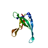
| ||||||||
|---|---|---|---|---|---|---|---|---|---|
| 1 | 
| ||||||||
| Unit cell |
|
-Components
| #1: Protein | Mass: 9683.214 Da / Num. of mol.: 1 Source method: isolated from a genetically manipulated source Source: (gene. exp.) Enterobacteria phage f1 (virus) / Genus: Inovirus / Species: Enterobacteria phage M13 / References: UniProt: P69543 |
|---|---|
| #2: Water | ChemComp-HOH /  Mass: 18.015 Da / Num. of mol.: 22 / Source method: isolated from a natural source / Formula: H2O Mass: 18.015 Da / Num. of mol.: 22 / Source method: isolated from a natural source / Formula: H2O |
-Experimental details
-Experiment
| Experiment | Method: X-RAY DIFFRACTION |
|---|
- Sample preparation
| Crystal | Density Matthews: 2.24 Å3/Da / Density % sol: 45.01 % | |||||||||||||||||||||||||
|---|---|---|---|---|---|---|---|---|---|---|---|---|---|---|---|---|---|---|---|---|---|---|---|---|---|---|
| Crystal grow | *PLUS pH: 7.5 / Method: vapor diffusion | |||||||||||||||||||||||||
| Components of the solutions | *PLUS
|
- Processing
| Refinement | Resolution: 2.2→8 Å / σ(F): 3
| ||||||||||||||||||||||||||||||||||||||||||||||||||||||||||||
|---|---|---|---|---|---|---|---|---|---|---|---|---|---|---|---|---|---|---|---|---|---|---|---|---|---|---|---|---|---|---|---|---|---|---|---|---|---|---|---|---|---|---|---|---|---|---|---|---|---|---|---|---|---|---|---|---|---|---|---|---|---|
| Refinement step | Cycle: LAST / Resolution: 2.2→8 Å
| ||||||||||||||||||||||||||||||||||||||||||||||||||||||||||||
| Refine LS restraints |
| ||||||||||||||||||||||||||||||||||||||||||||||||||||||||||||
| Refinement | *PLUS Rfactor Rfree: 0.172 / Rfactor Rwork: 0.18 | ||||||||||||||||||||||||||||||||||||||||||||||||||||||||||||
| Solvent computation | *PLUS | ||||||||||||||||||||||||||||||||||||||||||||||||||||||||||||
| Displacement parameters | *PLUS | ||||||||||||||||||||||||||||||||||||||||||||||||||||||||||||
| Refine LS restraints | *PLUS
|
