 Movie
Movie Controller

[English] 日本語
 Yorodumi
- PDB-1asw: AVIAN SARCOMA VIRUS INTEGRASE CATALYTIC CORE DOMAIN CRYSTALLIZED ... -
+ Open data

- Basic information
| Entry | Database: PDB / ID: 1asw | ||||||
|---|---|---|---|---|---|---|---|
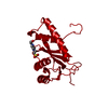
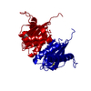
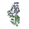
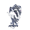
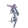
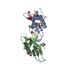
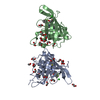
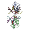
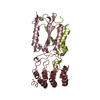
| Title | AVIAN SARCOMA VIRUS INTEGRASE CATALYTIC CORE DOMAIN CRYSTALLIZED FROM 20% PEG 4000, 10% ISOPROPANOL, HEPES PH 7.5 USING SELENOMETHIONINE SUBSTITUTED PROTEIN; DATA COLLECTED AT-165 DEGREES C | ||||||
 Components Components | AVIAN SARCOMA VIRUS INTEGRASE | ||||||
 Keywords Keywords | DNA INTEGRATION | ||||||
| Function / homology |  Function and homology information Function and homology informationHydrolases; Acting on peptide bonds (peptidases); Aspartic endopeptidases / ribonuclease H / DNA integration / viral genome integration into host DNA / establishment of integrated proviral latency / RNA-directed DNA polymerase / RNA stem-loop binding / virion component / RNA-directed DNA polymerase activity / RNA-DNA hybrid ribonuclease activity ...Hydrolases; Acting on peptide bonds (peptidases); Aspartic endopeptidases / ribonuclease H / DNA integration / viral genome integration into host DNA / establishment of integrated proviral latency / RNA-directed DNA polymerase / RNA stem-loop binding / virion component / RNA-directed DNA polymerase activity / RNA-DNA hybrid ribonuclease activity / Transferases; Transferring phosphorus-containing groups; Nucleotidyltransferases / viral nucleocapsid / DNA recombination / DNA-directed DNA polymerase / aspartic-type endopeptidase activity / Hydrolases; Acting on ester bonds / DNA-directed DNA polymerase activity / viral translational frameshifting / symbiont entry into host cell / proteolysis / DNA binding / zinc ion binding Similarity search - Function | ||||||
| Biological species |  Avian sarcoma virus Avian sarcoma virus | ||||||
| Method |  X-RAY DIFFRACTION / SYNCHROTRON / Resolution: 1.8 Å X-RAY DIFFRACTION / SYNCHROTRON / Resolution: 1.8 Å | ||||||
 Authors Authors | Bujacz, G. / Jaskolski, M. / Alexandratos, J. / Wlodawer, A. | ||||||
 Citation Citation | Journal: J.Mol.Biol. / Year: 1995 Title: High-resolution structure of the catalytic domain of avian sarcoma virus integrase. Authors: Bujacz, G. / Jaskolski, M. / Alexandratos, J. / Wlodawer, A. / Merkel, G. / Katz, R.A. / Skalka, A.M. #1: Journal: J.Biol.Chem. / Year: 1990Title: Expression, Purification, and Crystallization of Natural and Selenomethionyl Recombinant Ribonuclease H from Escherichia Coli Authors: Yang, W. / Hendrickson, W.A. / Kalman, E.T. / Crouch, R.J. | ||||||
| History |
|
- Structure visualization
| Structure viewer | Molecule: MolmilJmol/JSmol |
|---|
- Downloads & links
-Download
| PDBx/mmCIF format | 1asw.cif.gz | 49.5 KB | Display | PDBx/mmCIF format |
|---|---|---|---|---|
| PDB format | pdb1asw.ent.gz | 33.8 KB | Display | PDB format |
| PDBx/mmJSON format | 1asw.json.gz | Tree view | PDBx/mmJSON format | |
| Others |  Other downloads Other downloads |
-Validation report
| Arichive directory | https://data.pdbj.org/pub/pdb/validation_reports/as/1aswftp://data.pdbj.org/pub/pdb/validation_reports/as/1asw | HTTPS FTP |
|---|
-Related structure data


-Links
 PDBj
PDBj

- Assembly
| Deposited unit | 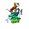
| ||||||||
|---|---|---|---|---|---|---|---|---|---|
| 1 | 
| ||||||||
| Unit cell |
| ||||||||
| Atom site foot note | 1: CIS PROLINE - PRO 73 |
-Components
| #1: Protein | Mass: 18083.082 Da / Num. of mol.: 1 Mutation: INS(PRO 48, LEU 49, ARG 50, GLU 51, ASN 208, LEU 209) Source method: isolated from a genetically manipulated source Details: CRYSTALLIZED FROM 20% PEG 4000, 10% ISOPROPANOL, HEPES PH 7.5 USING SELENOMETHIONINE-SUBSTITUTED PROTEIN, DATA COLLECTED AT LOW TEMPERATURE Source: (gene. exp.) Avian sarcoma virus / Genus: Alpharetrovirus / Strain: SCHMIDT-RUPPIN B / Plasmid: PRC23IN(52-207) / Production host:  |
|---|---|
| #2: Chemical | ChemComp-EPE /   Mass: 238.305 Da / Num. of mol.: 1 / Source method: obtained synthetically / Formula: C8H18N2O4S / Comment: pH buffer*YM Mass: 238.305 Da / Num. of mol.: 1 / Source method: obtained synthetically / Formula: C8H18N2O4S / Comment: pH buffer*YM |
| #3: Chemical | ChemComp-IPA /   Mass: 60.095 Da / Num. of mol.: 1 / Source method: obtained synthetically / Formula: C3H8O / Comment: alkaloid*YM Mass: 60.095 Da / Num. of mol.: 1 / Source method: obtained synthetically / Formula: C3H8O / Comment: alkaloid*YM |
| #4: Water | ChemComp-HOH /  Mass: 18.015 Da / Num. of mol.: 187 / Source method: isolated from a natural source / Formula: H2O Mass: 18.015 Da / Num. of mol.: 187 / Source method: isolated from a natural source / Formula: H2O |
| Has protein modification | Y |
| Source details | ORIGINAL VIRAL DNA CLONE: JU ET AL., J. VIROL. 33:1026-1033 (1980) ORIGINAL EXPRESSION CLONE: TERRY ...ORIGINAL VIRAL DNA CLONE: JU ET AL., J. VIROL. 33:1026-1033 (1980) ORIGINAL EXPRESSION |
-Experimental details
-Experiment
| Experiment | Method: X-RAY DIFFRACTION |
|---|
- Sample preparation
| Crystal | Density Matthews: 2.38 Å3/Da / Density % sol: 48.24 % | |||||||||||||||||||||||||
|---|---|---|---|---|---|---|---|---|---|---|---|---|---|---|---|---|---|---|---|---|---|---|---|---|---|---|
| Crystal grow | pH: 7.5 / Details: pH 7.5 | |||||||||||||||||||||||||
| Crystal grow | *PLUS Method: unknown | |||||||||||||||||||||||||
| Components of the solutions | *PLUS
|
-Data collection
| Diffraction | Mean temperature: 108 K | ||||||||||||
|---|---|---|---|---|---|---|---|---|---|---|---|---|---|
| Diffraction source | Source: SYNCHROTRON / Site: CHESS  / Beamline: F2 / Wavelength: 0.9464, 0.9792, 0.9790 / Beamline: F2 / Wavelength: 0.9464, 0.9792, 0.9790 | ||||||||||||
| Detector | Type: CUSTOM-MADE / Detector: CCD / Date: Feb 10, 1995 | ||||||||||||
| Radiation | Monochromatic (M) / Laue (L): M / Scattering type: x-ray | ||||||||||||
| Radiation wavelength |
| ||||||||||||
| Reflection | Resolution: 1.8→25 Å / Num. obs: 12157 / % possible obs: 94.2 % / Observed criterion σ(I): 0 / Redundancy: 7.07 % / Rmerge(I) obs: 0.068 | ||||||||||||
| Reflection | *PLUS Rmerge(I) obs: 0.068 |
- Processing
| Software |
| ||||||||||||||||||||||||||||||||||||||||||||||||||||||||||||||||||||||||||||||||||||
|---|---|---|---|---|---|---|---|---|---|---|---|---|---|---|---|---|---|---|---|---|---|---|---|---|---|---|---|---|---|---|---|---|---|---|---|---|---|---|---|---|---|---|---|---|---|---|---|---|---|---|---|---|---|---|---|---|---|---|---|---|---|---|---|---|---|---|---|---|---|---|---|---|---|---|---|---|---|---|---|---|---|---|---|---|---|
| Refinement | Resolution: 1.8→6 Å / σ(F): 2
| ||||||||||||||||||||||||||||||||||||||||||||||||||||||||||||||||||||||||||||||||||||
| Displacement parameters | Biso mean: 18.24 Å2 | ||||||||||||||||||||||||||||||||||||||||||||||||||||||||||||||||||||||||||||||||||||
| Refinement step | Cycle: LAST / Resolution: 1.8→6 Å
| ||||||||||||||||||||||||||||||||||||||||||||||||||||||||||||||||||||||||||||||||||||
| Refine LS restraints |
| ||||||||||||||||||||||||||||||||||||||||||||||||||||||||||||||||||||||||||||||||||||
| Software | *PLUS Name: PROLSQ / Classification: refinement | ||||||||||||||||||||||||||||||||||||||||||||||||||||||||||||||||||||||||||||||||||||
| Refinement | *PLUS % reflection Rfree: 8 % | ||||||||||||||||||||||||||||||||||||||||||||||||||||||||||||||||||||||||||||||||||||
| Solvent computation | *PLUS | ||||||||||||||||||||||||||||||||||||||||||||||||||||||||||||||||||||||||||||||||||||
| Displacement parameters | *PLUS |
