 Movie
Movie Controller

[English] 日本語
 Yorodumi
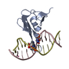
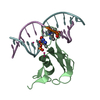
- PDB-6cc8: Crystal structure MBD3 MBD domain in complex with methylated CpG DNA -
+ Open data

- Basic information
| Entry | Database: PDB / ID: 6cc8 | |||||||||
|---|---|---|---|---|---|---|---|---|---|---|
| Title | Crystal structure MBD3 MBD domain in complex with methylated CpG DNA | |||||||||
 Components Components |
| |||||||||
 Keywords Keywords | DNA BINDING PROTEIN/DNA / Structural Genomics Consortium / SGC / DNA BINDING PROTEIN-DNA complex | |||||||||
| Function / homology |  Function and homology information Function and homology informationresponse to bisphenol A / ventricular cardiac muscle tissue development / NuRD complex / regulation of cell fate specification / regulation of stem cell differentiation / methyl-CpG binding / DNA methylation-dependent constitutive heterochromatin formation / RNA Polymerase I Transcription Initiation / embryonic organ development / Transcriptional regulation of brown and beige adipocyte differentiation by EBF2 ...response to bisphenol A / ventricular cardiac muscle tissue development / NuRD complex / regulation of cell fate specification / regulation of stem cell differentiation / methyl-CpG binding / DNA methylation-dependent constitutive heterochromatin formation / RNA Polymerase I Transcription Initiation / embryonic organ development / Transcriptional regulation of brown and beige adipocyte differentiation by EBF2 / Regulation of TP53 Activity through Acetylation / heterochromatin / Regulation of PTEN gene transcription / ERCC6 (CSB) and EHMT2 (G9a) positively regulate rRNA expression / Regulation of endogenous retroelements by KRAB-ZFP proteins / HDACs deacetylate histones / Regulation of endogenous retroelements by Piwi-interacting RNAs (piRNAs) / response to nutrient levels / response to estradiol / in utero embryonic development / Potential therapeutics for SARS / chromatin remodeling / negative regulation of DNA-templated transcription / positive regulation of DNA-templated transcription / chromatin / negative regulation of transcription by RNA polymerase II / protein-containing complex / DNA binding / nucleoplasm / nucleus / cytoplasm Similarity search - Function | |||||||||
| Biological species |  Homo sapiens (human) Homo sapiens (human)synthetic construct (others) | |||||||||
| Method |  X-RAY DIFFRACTION / SYNCHROTRON / MOLECULAR REPLACEMENT / Resolution: 1.95 Å X-RAY DIFFRACTION / SYNCHROTRON / MOLECULAR REPLACEMENT / Resolution: 1.95 Å | |||||||||
 Authors Authors | Liu, K. / Tempel, W. / Bountra, C. / Arrowsmith, C.H. / Edwards, A.M. / Min, J. / Structural Genomics Consortium (SGC) | |||||||||
 Citation Citation | Journal: Febs J. / Year: 2019 Title: Structural analyses reveal that MBD3 is a methylated CG binder. Authors: Liu, K. / Lei, M. / Wu, Z. / Gan, B. / Cheng, H. / Li, Y. / Min, J. | |||||||||
| History |
|
- Structure visualization
| Structure viewer | Molecule: MolmilJmol/JSmol |
|---|
- Downloads & links
-Download
| PDBx/mmCIF format | 6cc8.cif.gz | 124.2 KB | Display | PDBx/mmCIF format |
|---|---|---|---|---|
| PDB format | pdb6cc8.ent.gz | 93.6 KB | Display | PDB format |
| PDBx/mmJSON format | 6cc8.json.gz | Tree view | PDBx/mmJSON format | |
| Others |  Other downloads Other downloads |
-Validation report
| Arichive directory | https://data.pdbj.org/pub/pdb/validation_reports/cc/6cc8ftp://data.pdbj.org/pub/pdb/validation_reports/cc/6cc8 | HTTPS FTP |
|---|
-Related structure data
| Related structure data | 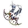 6ccgC 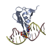 6ceuC  6cevC  6c2k S: Starting model for refinement C: citing same article ( |
|---|---|
| Similar structure data |


-Links
 PDBj
PDBj


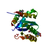
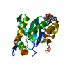
- Assembly
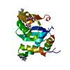
| Deposited unit | 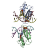
| ||||||||
|---|---|---|---|---|---|---|---|---|---|
| 1 | 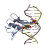
| ||||||||
| 2 | 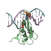
| ||||||||
| Unit cell |
|
-Components
| #1: Protein | Mass: 8473.716 Da / Num. of mol.: 2 / Fragment: MBD domain (UNP residues 1-71) Source method: isolated from a genetically manipulated source Source: (gene. exp.) Homo sapiens (human) / Gene: MBD3 / Plasmid: pET28-GSTLIC / Production host:  #2: DNA chain | Mass: 3677.419 Da / Num. of mol.: 4 / Source method: obtained synthetically / Details: dodecanucleotide / Source: (synth.) synthetic construct (others) #3: Chemical | ChemComp-UNX /   Mass: 103.120 Da / Num. of mol.: 12 / Source method: obtained synthetically Mass: 103.120 Da / Num. of mol.: 12 / Source method: obtained synthetically#4: Water | ChemComp-HOH / |  Mass: 18.015 Da / Num. of mol.: 64 / Source method: isolated from a natural source / Formula: H2O Mass: 18.015 Da / Num. of mol.: 64 / Source method: isolated from a natural source / Formula: H2O |
|---|
-Experimental details
-Experiment
| Experiment | Method: X-RAY DIFFRACTION / Number of used crystals: 1 |
|---|
- Sample preparation
| Crystal | Density Matthews: 2.8 Å3/Da / Density % sol: 55.4 % |
|---|---|
| Crystal grow | Temperature: 291 K / Method: vapor diffusion / pH: 7.5 Details: 25% PEG3350, 0.2 M sodium chloride, 0.1 M HEPES, 5% ethylene glycol |
-Data collection
| Diffraction | Mean temperature: 100 K |
|---|---|
| Diffraction source | Source: SYNCHROTRON / Site: APS  / Beamline: 19-ID / Wavelength: 0.97923 Å / Beamline: 19-ID / Wavelength: 0.97923 Å |
| Detector | Type: ADSC QUANTUM 315r / Detector: CCD / Date: Oct 20, 2012 |
| Radiation | Monochromator: double crystal Si(111) / Protocol: SINGLE WAVELENGTH / Monochromatic (M) / Laue (L): M / Scattering type: x-ray |
| Radiation wavelength | Wavelength: 0.97923 Å / Relative weight: 1 |
| Reflection | Resolution: 1.95→35.73 Å / Num. obs: 25160 / % possible obs: 99.9 % / Redundancy: 5.3 % / Rmerge(I) obs: 0.074 / Net I/σ(I): 14 |
| Reflection shell | Resolution: 1.95→2 Å / Redundancy: 3.7 % / Rmerge(I) obs: 0.761 / % possible all: 99.8 |
- Processing
| Software |
| ||||||||||||||||||||||||||||||||||||||||||||||||||||||||||||||||||||||||||||||||||||||||||||||||||||||||||||||||||||||||||||||||||||||||||||||||||||||||||||||||||||||||||||||||||||||
|---|---|---|---|---|---|---|---|---|---|---|---|---|---|---|---|---|---|---|---|---|---|---|---|---|---|---|---|---|---|---|---|---|---|---|---|---|---|---|---|---|---|---|---|---|---|---|---|---|---|---|---|---|---|---|---|---|---|---|---|---|---|---|---|---|---|---|---|---|---|---|---|---|---|---|---|---|---|---|---|---|---|---|---|---|---|---|---|---|---|---|---|---|---|---|---|---|---|---|---|---|---|---|---|---|---|---|---|---|---|---|---|---|---|---|---|---|---|---|---|---|---|---|---|---|---|---|---|---|---|---|---|---|---|---|---|---|---|---|---|---|---|---|---|---|---|---|---|---|---|---|---|---|---|---|---|---|---|---|---|---|---|---|---|---|---|---|---|---|---|---|---|---|---|---|---|---|---|---|---|---|---|---|---|
| Refinement | Method to determine structure: MOLECULAR REPLACEMENT Starting model: PDB ENTRY 6C2K 6c2k Resolution: 1.95→35.73 Å / Cor.coef. Fo:Fc: 0.949 / Cor.coef. Fo:Fc free: 0.935 / SU B: 9.689 / SU ML: 0.132 / Cross valid method: THROUGHOUT / σ(F): 0 / ESU R: 0.17 / ESU R Free: 0.156 Details: MODEL WAS ORIGINALLY REFINED AGAINST DATA COLLECTED ON AN ISOMORPHOUS CRYSTAL. THAT STRUCTURE WAS SOLVED BY MOLECULAR REPLACEMENT USING PHASER AND MOLREP TO POSITION FIRST AND SECOND COPY OF ...Details: MODEL WAS ORIGINALLY REFINED AGAINST DATA COLLECTED ON AN ISOMORPHOUS CRYSTAL. THAT STRUCTURE WAS SOLVED BY MOLECULAR REPLACEMENT USING PHASER AND MOLREP TO POSITION FIRST AND SECOND COPY OF THE COMPLEX, RESPECTIVELY. REFMAC WAS USED DURING INTERMEDIATE REFINEMENT. COOT WAS USED FOR INTERACTIVE MODEL BUILDING. MODEL GEOMETRY WAS ASSESSED ON THE MOLPROBITY SERVER.
| ||||||||||||||||||||||||||||||||||||||||||||||||||||||||||||||||||||||||||||||||||||||||||||||||||||||||||||||||||||||||||||||||||||||||||||||||||||||||||||||||||||||||||||||||||||||
| Solvent computation | Ion probe radii: 0.8 Å / Shrinkage radii: 0.8 Å / VDW probe radii: 1.2 Å | ||||||||||||||||||||||||||||||||||||||||||||||||||||||||||||||||||||||||||||||||||||||||||||||||||||||||||||||||||||||||||||||||||||||||||||||||||||||||||||||||||||||||||||||||||||||
| Displacement parameters | Biso mean: 42.99 Å2
| ||||||||||||||||||||||||||||||||||||||||||||||||||||||||||||||||||||||||||||||||||||||||||||||||||||||||||||||||||||||||||||||||||||||||||||||||||||||||||||||||||||||||||||||||||||||
| Refinement step | Cycle: LAST / Resolution: 1.95→35.73 Å
| ||||||||||||||||||||||||||||||||||||||||||||||||||||||||||||||||||||||||||||||||||||||||||||||||||||||||||||||||||||||||||||||||||||||||||||||||||||||||||||||||||||||||||||||||||||||
| Refine LS restraints |
|