 Movie
Movie Controller

[English] 日本語
 Yorodumi
- PDB-2vsc: Structure of the immunoglobulin-superfamily ectodomain of human CD47 -
+ Open data

- Basic information
| Entry | Database: PDB / ID: 2vsc | |||||||||
|---|---|---|---|---|---|---|---|---|---|---|
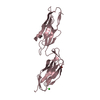
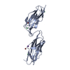
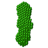
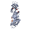
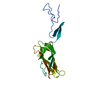
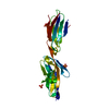
| Title | Structure of the immunoglobulin-superfamily ectodomain of human CD47 | |||||||||
 Components Components | LEUKOCYTE SURFACE ANTIGEN CD47 | |||||||||
 Keywords Keywords | CELL ADHESION / IMMUNOGLOBULIN DOMAIN / SIGNAL REGULATORY PROTEIN / CD47 / MEMBRANE / GLYCOPROTEIN / IMMUNOGLOBULIN SUPERFAMILY / PYRROLIDONE CARBOXYLIC ACID / TRANSMEMBRANE / PAIRED RECEPTOR / ALTERNATIVE SPLICING | |||||||||
| Function / homology |  Function and homology information Function and homology informationcellular response to interleukin-12 / regulation of Fc receptor mediated stimulatory signaling pathway / protein binding involved in heterotypic cell-cell adhesion / positive regulation of monocyte extravasation / regulation of interleukin-10 production / cell-cell adhesion mediator activity / positive regulation of cell-cell adhesion / regulation of type II interferon production / ATP export / fibrinogen binding ...cellular response to interleukin-12 / regulation of Fc receptor mediated stimulatory signaling pathway / protein binding involved in heterotypic cell-cell adhesion / positive regulation of monocyte extravasation / regulation of interleukin-10 production / cell-cell adhesion mediator activity / positive regulation of cell-cell adhesion / regulation of type II interferon production / ATP export / fibrinogen binding / regulation of tumor necrosis factor production / regulation of interleukin-12 production / regulation of nitric oxide biosynthetic process / regulation of interleukin-6 production / Signal regulatory protein family interactions / negative regulation of phagocytosis / thrombospondin receptor activity / tertiary granule membrane / cellular response to interleukin-1 / Integrin cell surface interactions / specific granule membrane / positive regulation of stress fiber assembly / positive regulation of phagocytosis / Cell surface interactions at the vascular wall / integrin-mediated signaling pathway / cellular response to type II interferon / positive regulation of T cell activation / positive regulation of inflammatory response / cell migration / angiogenesis / inflammatory response / apoptotic process / positive regulation of cell population proliferation / Neutrophil degranulation / cell surface / extracellular exosome / plasma membrane Similarity search - Function | |||||||||
| Biological species |  HOMO SAPIENS (human) HOMO SAPIENS (human) | |||||||||
| Method |  X-RAY DIFFRACTION / SYNCHROTRON / MOLECULAR REPLACEMENT / Resolution: 1.9 Å X-RAY DIFFRACTION / SYNCHROTRON / MOLECULAR REPLACEMENT / Resolution: 1.9 Å | |||||||||
 Authors Authors | Hatherley, D. / Graham, S.C. / Turner, J. / Harlos, K. / Stuart, D.I. / Barclay, A.N. | |||||||||
 Citation Citation | Journal: Mol.Cell / Year: 2008 Title: Paired Receptor Specificity Explained by Structures of Signal Regulatory Proteins Alone and Complexed with Cd47. Authors: Hatherley, D. / Graham, S.C. / Turner, J. / Harlos, K. / Stuart, D.I. / Barclay, A.N. | |||||||||
| History |
| |||||||||
| Remark 650 | HELIX DETERMINATION METHOD: AUTHOR PROVIDED. |
- Structure visualization
| Structure viewer | Molecule: MolmilJmol/JSmol |
|---|
- Downloads & links
-Download
| PDBx/mmCIF format | 2vsc.cif.gz | 212.7 KB | Display | PDBx/mmCIF format |
|---|---|---|---|---|
| PDB format | pdb2vsc.ent.gz | 173.6 KB | Display | PDB format |
| PDBx/mmJSON format | 2vsc.json.gz | Tree view | PDBx/mmJSON format | |
| Others |  Other downloads Other downloads |
-Validation report
| Arichive directory | https://data.pdbj.org/pub/pdb/validation_reports/vs/2vscftp://data.pdbj.org/pub/pdb/validation_reports/vs/2vsc | HTTPS FTP |
|---|
-Related structure data
| Related structure data | 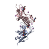 2jjsSC 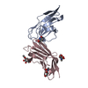 2jjtC 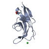 2jjuC 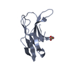 2jjvC 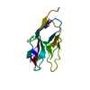 2jjwC S: Starting model for refinement C: citing same article ( |
|---|---|
| Similar structure data |


-Links
 PDBj
PDBj


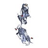
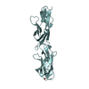
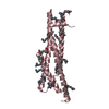
- Assembly
| Deposited unit | 
| ||||||||
|---|---|---|---|---|---|---|---|---|---|
| 1 | 
| ||||||||
| 2 | 
| ||||||||
| Unit cell |
| ||||||||
| Components on special symmetry positions |
|
-Components
| #1: Antibody | Mass: 14518.302 Da / Num. of mol.: 4 Fragment: IMMUNOGLOBULIN-SUPERFAMILY ECTODOMAIN, RESIDUES 19-136 Mutation: YES Source method: isolated from a genetically manipulated source Source: (gene. exp.) HOMO SAPIENS (human) / Plasmid: PEE14 / Cell line (production host): CHO / Production host:   CRICETULUS GRISEUS (Chinese hamster) / Variant (production host): LEC3.2.8.1 / References: UniProt: Q08722 CRICETULUS GRISEUS (Chinese hamster) / Variant (production host): LEC3.2.8.1 / References: UniProt: Q08722#2: Sugar | ChemComp-NAG /   Type: D-saccharide, beta linking / Mass: 221.208 Da / Num. of mol.: 12 Type: D-saccharide, beta linking / Mass: 221.208 Da / Num. of mol.: 12Source method: isolated from a genetically manipulated source Formula: C8H15NO6 #3: Chemical |   Mass: 24.305 Da / Num. of mol.: 2 / Source method: obtained synthetically / Formula: Mg Mass: 24.305 Da / Num. of mol.: 2 / Source method: obtained synthetically / Formula: Mg#4: Water | ChemComp-HOH / |  Mass: 18.015 Da / Num. of mol.: 353 / Source method: isolated from a natural source / Formula: H2O Mass: 18.015 Da / Num. of mol.: 353 / Source method: isolated from a natural source / Formula: H2OHas protein modification | Y | Sequence details | RESIDUE NUMBERING IS FOR MATURE PROTEIN (LACKING N- TERMINAL 18 AMINO ACID SIGNAL SEQUENCE). ...RESIDUE NUMBERING IS FOR MATURE PROTEIN (LACKING N- TERMINAL 18 AMINO ACID SIGNAL SEQUENCE). RESIDUE 1 (GLN) CYCLISES TO FORM A PYROGLUTAM | |
|---|
-Experimental details
-Experiment
| Experiment | Method: X-RAY DIFFRACTION / Number of used crystals: 1 |
|---|
- Sample preparation
| Crystal | Density Matthews: 2.6 Å3/Da / Density % sol: 52.5 % / Description: NONE |
|---|---|
| Crystal grow | pH: 5.5 Details: 200 NL 17 MG/ML CD47 PLUS 100 NL RESERVOIR (0.2 M MGCL2, 0.1 M BIS-TRIS PH 5.5, 25% W/V PEG 3350) EQUILIBRATED AGAINST 95 UL OF RESERVOIR AT 20.5 C. |
-Data collection
| Diffraction | Mean temperature: 100 K |
|---|---|
| Diffraction source | Source: SYNCHROTRON / Site: ESRF  / Beamline: BM14 / Wavelength: 0.978 / Beamline: BM14 / Wavelength: 0.978 |
| Detector | Type: MARRESEARCH / Detector: CCD / Date: Apr 12, 2006 / Details: MIRRORS |
| Radiation | Monochromator: SI(111) MONOCHROMATOR / Protocol: SINGLE WAVELENGTH / Monochromatic (M) / Laue (L): M / Scattering type: x-ray |
| Radiation wavelength | Wavelength: 0.978 Å / Relative weight: 1 |
| Reflection | Resolution: 1.9→28.3 Å / Num. obs: 41496 / % possible obs: 98.8 % / Observed criterion σ(I): -3 / Redundancy: 3.7 % / Biso Wilson estimate: 24.9 Å2 / Rmerge(I) obs: 0.07 / Net I/σ(I): 13.9 |
| Reflection shell | Resolution: 1.9→2 Å / Redundancy: 3.1 % / Rmerge(I) obs: 0.4 / Mean I/σ(I) obs: 2.9 / % possible all: 91.8 |
- Processing
| Software |
| ||||||||||||||||||||||||||||||||||||||||||||||||||||||||||||||||||||||||||||||||||||||||||||||||||||||||||||||||||||||||||||||||||||||||||||||||||||||||||||||||||||||||||||||||||||||
|---|---|---|---|---|---|---|---|---|---|---|---|---|---|---|---|---|---|---|---|---|---|---|---|---|---|---|---|---|---|---|---|---|---|---|---|---|---|---|---|---|---|---|---|---|---|---|---|---|---|---|---|---|---|---|---|---|---|---|---|---|---|---|---|---|---|---|---|---|---|---|---|---|---|---|---|---|---|---|---|---|---|---|---|---|---|---|---|---|---|---|---|---|---|---|---|---|---|---|---|---|---|---|---|---|---|---|---|---|---|---|---|---|---|---|---|---|---|---|---|---|---|---|---|---|---|---|---|---|---|---|---|---|---|---|---|---|---|---|---|---|---|---|---|---|---|---|---|---|---|---|---|---|---|---|---|---|---|---|---|---|---|---|---|---|---|---|---|---|---|---|---|---|---|---|---|---|---|---|---|---|---|---|---|
| Refinement | Method to determine structure: MOLECULAR REPLACEMENT Starting model: PDB ENTRY 2JJS, CHAIN C Resolution: 1.9→28.19 Å / Cor.coef. Fo:Fc: 0.938 / Cor.coef. Fo:Fc free: 0.909 / SU B: 8.331 / SU ML: 0.135 / Cross valid method: THROUGHOUT / ESU R: 0.18 / ESU R Free: 0.166 / Stereochemistry target values: MAXIMUM LIKELIHOOD Details: HYDROGENS HAVE BEEN ADDED IN THE RIDING POSITIONS. B VALUES INCLUDE TLS CONTRIBUTIONS.
| ||||||||||||||||||||||||||||||||||||||||||||||||||||||||||||||||||||||||||||||||||||||||||||||||||||||||||||||||||||||||||||||||||||||||||||||||||||||||||||||||||||||||||||||||||||||
| Solvent computation | Ion probe radii: 0.8 Å / Shrinkage radii: 0.8 Å / VDW probe radii: 1.2 Å / Solvent model: MASK | ||||||||||||||||||||||||||||||||||||||||||||||||||||||||||||||||||||||||||||||||||||||||||||||||||||||||||||||||||||||||||||||||||||||||||||||||||||||||||||||||||||||||||||||||||||||
| Displacement parameters | Biso mean: 25.59 Å2
| ||||||||||||||||||||||||||||||||||||||||||||||||||||||||||||||||||||||||||||||||||||||||||||||||||||||||||||||||||||||||||||||||||||||||||||||||||||||||||||||||||||||||||||||||||||||
| Refinement step | Cycle: LAST / Resolution: 1.9→28.19 Å
| ||||||||||||||||||||||||||||||||||||||||||||||||||||||||||||||||||||||||||||||||||||||||||||||||||||||||||||||||||||||||||||||||||||||||||||||||||||||||||||||||||||||||||||||||||||||
| Refine LS restraints |
|