 Movie
Movie Controller


+ Open data

- Basic information
| Entry | Database: PDB / ID: 2jjv | ||||||
|---|---|---|---|---|---|---|---|
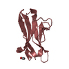
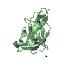
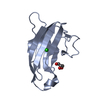
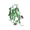
| Title | Structure of human signal regulatory protein (sirp) beta(2) | ||||||
 Components Components | SIGNAL-REGULATORY PROTEIN BETA 1. | ||||||
 Keywords Keywords | IMMUNE SYSTEM / IMMUNOGLOBULIN SUPERFAMILY / SIGNAL REGULATORY PROTEIN BETA / SIRP / SIRPB2 / PAIRED RECEPTOR | ||||||
| Function / homology |  Function and homology information Function and homology information | ||||||
| Biological species |  HOMO SAPIENS (human) HOMO SAPIENS (human) | ||||||
| Method |  X-RAY DIFFRACTION / SYNCHROTRON / MOLECULAR REPLACEMENT / Resolution: 1.8 Å X-RAY DIFFRACTION / SYNCHROTRON / MOLECULAR REPLACEMENT / Resolution: 1.8 Å | ||||||
 Authors Authors | Hatherley, D. / Graham, S.C. / Turner, J. / Harlos, K. / Stuart, D.I. / Barclay, A.N. | ||||||
 Citation Citation | Journal: Mol.Cell / Year: 2008 Title: Paired Receptor Specificity Explained by Structures of Signal Regulatory Proteins Alone and Complexed with Cd47. Authors: Hatherley, D. / Graham, S.C. / Turner, J. / Harlos, K. / Stuart, D.I. / Barclay, A.N. | ||||||
| History |
| ||||||
| Remark 650 | HELIX DETERMINATION METHOD: AUTHOR PROVIDED. | ||||||
| Remark 700 | SHEET THE SHEET STRUCTURE OF THIS MOLECULE IS BIFURCATED. IN ORDER TO REPRESENT THIS FEATURE IN ... SHEET THE SHEET STRUCTURE OF THIS MOLECULE IS BIFURCATED. IN ORDER TO REPRESENT THIS FEATURE IN THE SHEET RECORDS BELOW, TWO SHEETS ARE DEFINED. |
- Structure visualization
| Structure viewer | Molecule: MolmilJmol/JSmol |
|---|
- Downloads & links
-Download
| PDBx/mmCIF format | 2jjv.cif.gz | 105.2 KB | Display | PDBx/mmCIF format |
|---|---|---|---|---|
| PDB format | pdb2jjv.ent.gz | 82.1 KB | Display | PDB format |
| PDBx/mmJSON format | 2jjv.json.gz | Tree view | PDBx/mmJSON format | |
| Others |  Other downloads Other downloads |
-Validation report
| Arichive directory | https://data.pdbj.org/pub/pdb/validation_reports/jj/2jjvftp://data.pdbj.org/pub/pdb/validation_reports/jj/2jjv | HTTPS FTP |
|---|
-Related structure data
| Related structure data | 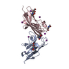 2jjsC 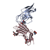 2jjtC 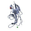 2jjuC 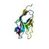 2jjwC 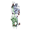 2vscC 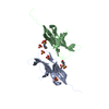 2uv3S C: citing same article ( S: Starting model for refinement |
|---|---|
| Similar structure data |


-Links
 PDBj
PDBj

- Assembly
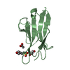
| Deposited unit | 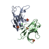
| ||||||||
|---|---|---|---|---|---|---|---|---|---|
| 1 | 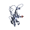
| ||||||||
| 2 | 
| ||||||||
| Unit cell |
|
-Components
| #1: Antibody | Mass: 14110.844 Da / Num. of mol.: 2 / Fragment: N-TERMINAL ECTODOMAIN, RESIDUES 30-148 Source method: isolated from a genetically manipulated source Details: 2 ISOFORMS PRODUCED BY ALTERNATIVE SPLICING. IN THIS ENTRY ISOFORM 1 (O00241-1) IS PRESENT Source: (gene. exp.) HOMO SAPIENS (human) / Plasmid: PEE14 / Cell line (production host): CHO / Production host:   CRICETULUS GRISEUS (Chinese hamster) / Variant (production host): LEC3.2.8.1 / References: UniProt: Q5TFQ8 CRICETULUS GRISEUS (Chinese hamster) / Variant (production host): LEC3.2.8.1 / References: UniProt: Q5TFQ8#2: Chemical | ChemComp-SO4 /   Mass: 96.063 Da / Num. of mol.: 4 / Source method: obtained synthetically / Formula: SO4 Mass: 96.063 Da / Num. of mol.: 4 / Source method: obtained synthetically / Formula: SO4#3: Chemical | ChemComp-CL / |   Mass: 35.453 Da / Num. of mol.: 1 / Source method: obtained synthetically / Formula: Cl Mass: 35.453 Da / Num. of mol.: 1 / Source method: obtained synthetically / Formula: Cl#4: Water | ChemComp-HOH / |  Mass: 18.015 Da / Num. of mol.: 139 / Source method: isolated from a natural source / Formula: H2O Mass: 18.015 Da / Num. of mol.: 139 / Source method: isolated from a natural source / Formula: H2OHas protein modification | Y | Sequence details | RESIDUE NUMBERING IS FOR THE MATURE PROTEIN (LACKING N- TERMINAL 29 AMINO ACID SIGNAL SEQUENCE). C- ...RESIDUE NUMBERING IS FOR THE MATURE PROTEIN (LACKING N- TERMINAL 29 AMINO ACID SIGNAL SEQUENCE). C-TERMINAL PURIFICATI | |
|---|
-Experimental details
-Experiment
| Experiment | Method: X-RAY DIFFRACTION / Number of used crystals: 1 |
|---|
- Sample preparation
| Crystal | Density Matthews: 1.94 Å3/Da / Density % sol: 36.5 % / Description: NONE |
|---|---|
| Crystal grow | pH: 6 Details: 300 NL 25 MG/ML SIRP BETA PLUS 100 NL RESERVOIR (3.2 M AMMONIUM SULPHATE, 0.1 M MES PH 6.0) EQUILIBRATED AGAINST 95 UL OF RESERVOIR AT 20.5 C. |
-Data collection
| Diffraction | Mean temperature: 100 K |
|---|---|
| Diffraction source | Source: SYNCHROTRON / Site: ESRF  / Beamline: BM14 / Wavelength: 0.979 / Beamline: BM14 / Wavelength: 0.979 |
| Detector | Type: MARRESEARCH / Detector: CCD / Date: Jun 16, 2007 / Details: MIRRORS |
| Radiation | Monochromator: SI(111) MONOCHROMATOR / Protocol: SINGLE WAVELENGTH / Monochromatic (M) / Laue (L): M / Scattering type: x-ray |
| Radiation wavelength | Wavelength: 0.979 Å / Relative weight: 1 |
| Reflection | Resolution: 1.8→50 Å / Num. obs: 19509 / % possible obs: 96.8 % / Observed criterion σ(I): -3 / Redundancy: 5.9 % / Biso Wilson estimate: 15.42 Å2 / Rmerge(I) obs: 0.07 / Net I/σ(I): 30 |
| Reflection shell | Resolution: 1.8→1.86 Å / Redundancy: 2.7 % / Rmerge(I) obs: 0.17 / Mean I/σ(I) obs: 4.4 / % possible all: 79.1 |
- Processing
| Software |
| ||||||||||||||||||||||||||||||||||||||||||||||||||||||||||||||||||||||||||||||||||||||||||||||||||||||||||||||||||||||||||||||||||||||||||||||||||||||||||||||||||||||||||||||||||||||
|---|---|---|---|---|---|---|---|---|---|---|---|---|---|---|---|---|---|---|---|---|---|---|---|---|---|---|---|---|---|---|---|---|---|---|---|---|---|---|---|---|---|---|---|---|---|---|---|---|---|---|---|---|---|---|---|---|---|---|---|---|---|---|---|---|---|---|---|---|---|---|---|---|---|---|---|---|---|---|---|---|---|---|---|---|---|---|---|---|---|---|---|---|---|---|---|---|---|---|---|---|---|---|---|---|---|---|---|---|---|---|---|---|---|---|---|---|---|---|---|---|---|---|---|---|---|---|---|---|---|---|---|---|---|---|---|---|---|---|---|---|---|---|---|---|---|---|---|---|---|---|---|---|---|---|---|---|---|---|---|---|---|---|---|---|---|---|---|---|---|---|---|---|---|---|---|---|---|---|---|---|---|---|---|
| Refinement | Method to determine structure: MOLECULAR REPLACEMENT Starting model: PDB ENTRY 2UV3 CHAIN A Resolution: 1.8→57.35 Å / Cor.coef. Fo:Fc: 0.952 / Cor.coef. Fo:Fc free: 0.929 / SU B: 4.741 / SU ML: 0.079 / Cross valid method: THROUGHOUT / ESU R: 0.135 / ESU R Free: 0.128 / Stereochemistry target values: MAXIMUM LIKELIHOOD Details: HYDROGENS HAVE BEEN ADDED IN THE RIDING POSITIONS. B VALUES INCLUDE TLS CONTRIBUTIONS.
| ||||||||||||||||||||||||||||||||||||||||||||||||||||||||||||||||||||||||||||||||||||||||||||||||||||||||||||||||||||||||||||||||||||||||||||||||||||||||||||||||||||||||||||||||||||||
| Solvent computation | Ion probe radii: 0.8 Å / Shrinkage radii: 0.8 Å / VDW probe radii: 1.2 Å / Solvent model: MASK | ||||||||||||||||||||||||||||||||||||||||||||||||||||||||||||||||||||||||||||||||||||||||||||||||||||||||||||||||||||||||||||||||||||||||||||||||||||||||||||||||||||||||||||||||||||||
| Displacement parameters | Biso mean: 10.99 Å2
| ||||||||||||||||||||||||||||||||||||||||||||||||||||||||||||||||||||||||||||||||||||||||||||||||||||||||||||||||||||||||||||||||||||||||||||||||||||||||||||||||||||||||||||||||||||||
| Refinement step | Cycle: LAST / Resolution: 1.8→57.35 Å
| ||||||||||||||||||||||||||||||||||||||||||||||||||||||||||||||||||||||||||||||||||||||||||||||||||||||||||||||||||||||||||||||||||||||||||||||||||||||||||||||||||||||||||||||||||||||
| Refine LS restraints |
|