 Movie
Movie Controller

[English] 日本語
 Yorodumi
- PDB-2v5q: CRYSTAL STRUCTURE OF WILD-TYPE PLK-1 KINASE DOMAIN IN COMPLEX WIT... -
+ Open data

- Basic information
| Entry | Database: PDB / ID: 2v5q | ||||||
|---|---|---|---|---|---|---|---|
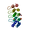
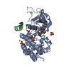
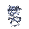
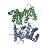
| Title | CRYSTAL STRUCTURE OF WILD-TYPE PLK-1 KINASE DOMAIN IN COMPLEX WITH A SELECTIVE DARPIN | ||||||
 Components Components |
| ||||||
 Keywords Keywords | TRANSFERASE / DESIGN ANKYRIN REPEAT PROTEIN / TRANSFERASE COMPLEX / PHOSPHORYLATION / NUCLEOTIDE-BINDING / SERINE/THREONINE-PROTEIN KINASE / KINASE / NUCLEUS / ATP-BINDING / SERINE/THREONINE PROTEIN KINASE | ||||||
| Function / homology |  Function and homology information Function and homology informationMitotic Telophase/Cytokinesis / regulation of protein localization to cell cortex / Mitotic Metaphase/Anaphase Transition / synaptonemal complex disassembly / Activation of NIMA Kinases NEK9, NEK6, NEK7 / polo kinase / mitotic nuclear membrane disassembly / Phosphorylation of Emi1 / protein localization to nuclear envelope / homologous chromosome segregation ...Mitotic Telophase/Cytokinesis / regulation of protein localization to cell cortex / Mitotic Metaphase/Anaphase Transition / synaptonemal complex disassembly / Activation of NIMA Kinases NEK9, NEK6, NEK7 / polo kinase / mitotic nuclear membrane disassembly / Phosphorylation of Emi1 / protein localization to nuclear envelope / homologous chromosome segregation / metaphase/anaphase transition of mitotic cell cycle / female meiosis chromosome segregation / nuclear membrane disassembly / synaptonemal complex / Phosphorylation of the APC/C / anaphase-promoting complex binding / Golgi inheritance / outer kinetochore / positive regulation of ubiquitin protein ligase activity / microtubule bundle formation / double-strand break repair via alternative nonhomologous end joining / mitotic chromosome condensation / Polo-like kinase mediated events / Golgi Cisternae Pericentriolar Stack Reorganization / regulation of mitotic spindle assembly / centrosome cycle / regulation of mitotic metaphase/anaphase transition / sister chromatid cohesion / positive regulation of ubiquitin-protein transferase activity / regulation of mitotic cell cycle phase transition / mitotic spindle assembly checkpoint signaling / mitotic spindle pole / spindle midzone / mitotic G2 DNA damage checkpoint signaling / regulation of anaphase-promoting complex-dependent catabolic process / mitotic cytokinesis / establishment of mitotic spindle orientation / mitotic sister chromatid segregation / positive regulation of proteolysis / Regulation of MITF-M-dependent genes involved in cell cycle and proliferation / negative regulation of double-strand break repair via homologous recombination / Cyclin A/B1/B2 associated events during G2/M transition / protein localization to chromatin / Amplification of signal from unattached kinetochores via a MAD2 inhibitory signal / Loss of Nlp from mitotic centrosomes / Loss of proteins required for interphase microtubule organization from the centrosome / Recruitment of mitotic centrosome proteins and complexes / centriole / Mitotic Prometaphase / Recruitment of NuMA to mitotic centrosomes / Anchoring of the basal body to the plasma membrane / EML4 and NUDC in mitotic spindle formation / regulation of mitotic cell cycle / AURKA Activation by TPX2 / Resolution of Sister Chromatid Cohesion / Condensation of Prophase Chromosomes / mitotic spindle organization / regulation of cytokinesis / establishment of protein localization / peptidyl-serine phosphorylation / RHO GTPases Activate Formins / APC/C:Cdh1 mediated degradation of Cdc20 and other APC/C:Cdh1 targeted proteins in late mitosis/early G1 / kinetochore / positive regulation of protein localization to nucleus / protein destabilization / G2/M transition of mitotic cell cycle / centriolar satellite / spindle / spindle pole / The role of GTSE1 in G2/M progression after G2 checkpoint / Separation of Sister Chromatids / Regulation of PLK1 Activity at G2/M Transition / mitotic cell cycle / double-strand break repair / positive regulation of proteasomal ubiquitin-dependent protein catabolic process / microtubule cytoskeleton / midbody / microtubule binding / protein phosphorylation / protein kinase activity / regulation of cell cycle / protein ubiquitination / protein serine kinase activity / protein serine/threonine kinase activity / centrosome / protein kinase binding / negative regulation of apoptotic process / chromatin / magnesium ion binding / negative regulation of transcription by RNA polymerase II / nucleoplasm / ATP binding / identical protein binding / nucleus / cytoplasm / cytosol Similarity search - Function | ||||||
| Biological species |  HOMO SAPIENS (human) HOMO SAPIENS (human)synthetic construct (others) | ||||||
| Method |  X-RAY DIFFRACTION / SYNCHROTRON / MOLECULAR REPLACEMENT / Resolution: 2.3 Å X-RAY DIFFRACTION / SYNCHROTRON / MOLECULAR REPLACEMENT / Resolution: 2.3 Å | ||||||
 Authors Authors | Bandeiras, T.M. / Hillig, R.C. / Matias, P.M. / Eberspaecher, U. / Fanghaenel, J. / Thomaz, M. / Miranda, S. / Crusius, K. / Puetter, V. / Amstutz, P. ...Bandeiras, T.M. / Hillig, R.C. / Matias, P.M. / Eberspaecher, U. / Fanghaenel, J. / Thomaz, M. / Miranda, S. / Crusius, K. / Puetter, V. / Amstutz, P. / Gulotti-Georgieva, M. / Binz, H.K. / Holz, C. / Schmitz, A.A.P. / Lang, C. / Donner, P. / Egner, U. / Carrondo, M.A. / Mueller-Tiemann, B. | ||||||
 Citation Citation | Journal: Acta Crystallogr. D Biol. Crystallogr. / Year: 2008 Title: Structure of wild-type Plk-1 kinase domain in complex with a selective DARPin. Authors: Bandeiras, T.M. / Hillig, R.C. / Matias, P.M. / Eberspaecher, U. / Fanghanel, J. / Thomaz, M. / Miranda, S. / Crusius, K. / Putter, V. / Amstutz, P. / Gulotti-Georgieva, M. / Binz, H.K. / ...Authors: Bandeiras, T.M. / Hillig, R.C. / Matias, P.M. / Eberspaecher, U. / Fanghanel, J. / Thomaz, M. / Miranda, S. / Crusius, K. / Putter, V. / Amstutz, P. / Gulotti-Georgieva, M. / Binz, H.K. / Holz, C. / Schmitz, A.A. / Lang, C. / Donner, P. / Egner, U. / Carrondo, M.A. / Muller-Tiemann, B. | ||||||
| History |
|
- Structure visualization
| Structure viewer | Molecule: MolmilJmol/JSmol |
|---|
- Downloads & links
-Download
| PDBx/mmCIF format | 2v5q.cif.gz | 180.8 KB | Display | PDBx/mmCIF format |
|---|---|---|---|---|
| PDB format | pdb2v5q.ent.gz | 142.7 KB | Display | PDB format |
| PDBx/mmJSON format | 2v5q.json.gz | Tree view | PDBx/mmJSON format | |
| Others |  Other downloads Other downloads |
-Validation report
| Arichive directory | https://data.pdbj.org/pub/pdb/validation_reports/v5/2v5qftp://data.pdbj.org/pub/pdb/validation_reports/v5/2v5q | HTTPS FTP |
|---|
-Related structure data


-Links
 PDBj
PDBj


- Assembly
| Deposited unit | 
| ||||||||
|---|---|---|---|---|---|---|---|---|---|
| 1 | 
| ||||||||
| 2 | 
| ||||||||
| Unit cell |
|
-Components
| #1: Protein | Mass: 35972.875 Da / Num. of mol.: 2 / Fragment: KINASE DOMAIN, RESIDUES 33-345 Source method: isolated from a genetically manipulated source Details: CONSTRUCT 4 / Source: (gene. exp.) HOMO SAPIENS (human) / Production host:   SPODOPTERA FRUGIPERDA (fall armyworm) / References: UniProt: P53350, polo kinase SPODOPTERA FRUGIPERDA (fall armyworm) / References: UniProt: P53350, polo kinase#2: Protein | Mass: 17835.967 Da / Num. of mol.: 2 Source method: isolated from a genetically manipulated source Details: VARIANT 3H10 / Source: (gene. exp.) synthetic construct (others) / Description: DESIGNED PROTEIN / Production host:  #3: Water | ChemComp-HOH / |  Mass: 18.015 Da / Num. of mol.: 406 / Source method: isolated from a natural source / Formula: H2O Mass: 18.015 Da / Num. of mol.: 406 / Source method: isolated from a natural source / Formula: H2O |
|---|
-Experimental details
-Experiment
| Experiment | Method: X-RAY DIFFRACTION / Number of used crystals: 1 |
|---|
- Sample preparation
| Crystal | Density Matthews: 2.7 Å3/Da / Density % sol: 54 % |
|---|---|
| Crystal grow | Temperature: 303 K Details: 0.1 M TRIS-HCL PH 8.0, 8% PEG 5000 MME, 0.01 M EDTA SODIUM SALT AT 303 K. |
-Data collection
| Diffraction | Mean temperature: 100 K |
|---|---|
| Diffraction source | Source: SYNCHROTRON / Site: ESRF  / Beamline: ID29 / Wavelength: 1.037 / Beamline: ID29 / Wavelength: 1.037 |
| Detector | Type: ADSC CCD / Detector: CCD / Date: Jul 20, 2006 / Details: MIRRORS |
| Radiation | Protocol: SINGLE WAVELENGTH / Monochromatic (M) / Laue (L): M / Scattering type: x-ray |
| Radiation wavelength | Wavelength: 1.037 Å / Relative weight: 1 |
| Reflection | Resolution: 2.3→96.2 Å / Num. obs: 51702 / % possible obs: 99.3 % / Redundancy: 3.5 % / Biso Wilson estimate: 45.7 Å2 / Rmerge(I) obs: 0.06 / Net I/σ(I): 15.7 |
| Reflection shell | Resolution: 2.3→2.42 Å / Redundancy: 3.6 % / Rmerge(I) obs: 0.4 / Mean I/σ(I) obs: 3.3 / % possible all: 99.3 |
- Processing
| Software |
| ||||||||||||||||||||||||||||||||||||||||||||||||||||||||||||||||||||||||||||||||||||||||||||||||||||||||||||||||||||||||||||||||||||||||||||||||||||||||||||||||||||||||||||||||||||||
|---|---|---|---|---|---|---|---|---|---|---|---|---|---|---|---|---|---|---|---|---|---|---|---|---|---|---|---|---|---|---|---|---|---|---|---|---|---|---|---|---|---|---|---|---|---|---|---|---|---|---|---|---|---|---|---|---|---|---|---|---|---|---|---|---|---|---|---|---|---|---|---|---|---|---|---|---|---|---|---|---|---|---|---|---|---|---|---|---|---|---|---|---|---|---|---|---|---|---|---|---|---|---|---|---|---|---|---|---|---|---|---|---|---|---|---|---|---|---|---|---|---|---|---|---|---|---|---|---|---|---|---|---|---|---|---|---|---|---|---|---|---|---|---|---|---|---|---|---|---|---|---|---|---|---|---|---|---|---|---|---|---|---|---|---|---|---|---|---|---|---|---|---|---|---|---|---|---|---|---|---|---|---|---|
| Refinement | Method to determine structure: MOLECULAR REPLACEMENT Starting model: PLK-1 - HOMOLOGY MODEL FROM PDB ENTRY 1OL5 DARPIN 3H10 - PDB ENTRY 1MJ0 TRUNCATED AFTER RESIDUE 141 Resolution: 2.3→61.08 Å / Cor.coef. Fo:Fc: 0.95 / Cor.coef. Fo:Fc free: 0.928 / SU B: 11.227 / SU ML: 0.145 / TLS residual ADP flag: LIKELY RESIDUAL / Cross valid method: THROUGHOUT / ESU R: 0.245 / ESU R Free: 0.198 / Stereochemistry target values: MAXIMUM LIKELIHOOD / Details: HYDROGENS HAVE BEEN ADDED IN THE RIDING POSITIONS.
| ||||||||||||||||||||||||||||||||||||||||||||||||||||||||||||||||||||||||||||||||||||||||||||||||||||||||||||||||||||||||||||||||||||||||||||||||||||||||||||||||||||||||||||||||||||||
| Solvent computation | Ion probe radii: 0.8 Å / Shrinkage radii: 0.8 Å / VDW probe radii: 1.4 Å / Solvent model: BABINET MODEL WITH MASK | ||||||||||||||||||||||||||||||||||||||||||||||||||||||||||||||||||||||||||||||||||||||||||||||||||||||||||||||||||||||||||||||||||||||||||||||||||||||||||||||||||||||||||||||||||||||
| Displacement parameters | Biso mean: 34.28 Å2
| ||||||||||||||||||||||||||||||||||||||||||||||||||||||||||||||||||||||||||||||||||||||||||||||||||||||||||||||||||||||||||||||||||||||||||||||||||||||||||||||||||||||||||||||||||||||
| Refinement step | Cycle: LAST / Resolution: 2.3→61.08 Å
| ||||||||||||||||||||||||||||||||||||||||||||||||||||||||||||||||||||||||||||||||||||||||||||||||||||||||||||||||||||||||||||||||||||||||||||||||||||||||||||||||||||||||||||||||||||||
| Refine LS restraints |
|