 Movie
Movie Controller

[English] 日本語
 Yorodumi
- PDB-2v5l: Structures of the Open and Closed State of Trypanosomal Triosepho... -
+ Open data

- Basic information
| Entry | Database: PDB / ID: 2v5l | ||||||
|---|---|---|---|---|---|---|---|
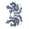
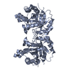
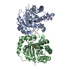
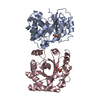
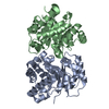
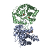
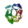
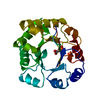
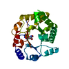
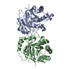
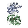
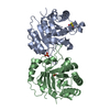
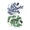
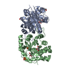
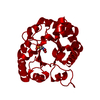
| Title | Structures of the Open and Closed State of Trypanosomal Triosephosphate Isomerase: as Observed in a New Crystal Form: Implications for the Reaction Mechanism | ||||||
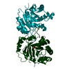
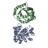
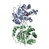
 Components Components | TRIOSEPHOSPHATE ISOMERASE | ||||||
 Keywords Keywords | OXIDOREDUCTASE / TRIOSEPHOSPHATE ISOMERASE / PENTOSE SHUNT / GLUCONEOGENESIS / BINDING STUDIES / TIM / ISOMERASE / GLYCOSOME / GLYCOLYSIS / ENGINEERING / LIPID SYNTHESIS / FATTY ACID BIOSYNTHESIS | ||||||
| Function / homology |  Function and homology informationglycosome / triose-phosphate isomerase / triose-phosphate isomerase activity / gluconeogenesis / glycolytic process / cytoplasm Function and homology informationglycosome / triose-phosphate isomerase / triose-phosphate isomerase activity / gluconeogenesis / glycolytic process / cytoplasmSimilarity search - Function | ||||||
| Biological species |  TRYPANOSOMA BRUCEI BRUCEI (eukaryote) TRYPANOSOMA BRUCEI BRUCEI (eukaryote) | ||||||
| Method | X-RAY DIFFRACTION / MOLECULAR REPLACEMENT / Resolution: 2.4 Å | ||||||
 Authors Authors | Noble, M.E.M. / Zeelen, J.P. / Wierenga, R.K. | ||||||
 Citation Citation | Journal: Proteins: Struct.,Funct., Genet. / Year: 1993 Title: Structures of the Open and Closed State of Trypanosomal Triosephosphate Isomerase: As Observed in a New Crystal Form: Implications for the Reaction Mechanism Authors: Noble, M.E.M. / Zeelen, J.P. / Wierenga, R.K. | ||||||
| History |
| ||||||
| Remark 700 | SHEET DETERMINATION METHOD: DSSP THE SHEETS PRESENTED AS "AA" IN EACH CHAIN ON SHEET RECORDS BELOW ... SHEET DETERMINATION METHOD: DSSP THE SHEETS PRESENTED AS "AA" IN EACH CHAIN ON SHEET RECORDS BELOW IS ACTUALLY AN 9-STRANDED BARREL THIS IS REPRESENTED BY A 10-STRANDED SHEET IN WHICH THE FIRST AND LAST STRANDS ARE IDENTICAL. |
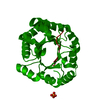
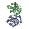
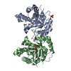
- Structure visualization
| Structure viewer | Molecule: MolmilJmol/JSmol |
|---|
- Downloads & links
-Download
| PDBx/mmCIF format | 2v5l.cif.gz | 104.1 KB | Display | PDBx/mmCIF format |
|---|---|---|---|---|
| PDB format | pdb2v5l.ent.gz | 84.4 KB | Display | PDB format |
| PDBx/mmJSON format | 2v5l.json.gz | Tree view | PDBx/mmJSON format | |
| Others |  Other downloads Other downloads |
-Validation report
| Arichive directory | https://data.pdbj.org/pub/pdb/validation_reports/v5/2v5lftp://data.pdbj.org/pub/pdb/validation_reports/v5/2v5l | HTTPS FTP |
|---|
-Related structure data


-Links
 PDBj
PDBj
- Assembly
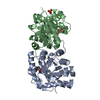
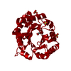
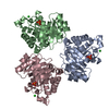
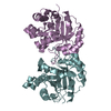
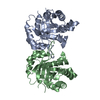
| Deposited unit | 
| ||||||||
|---|---|---|---|---|---|---|---|---|---|
| 1 | 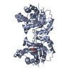
| ||||||||
| 2 | 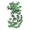
| ||||||||
| Unit cell |
| ||||||||
| Components on special symmetry positions |
|
-Components
| #1: Protein | / TRIOSE-PHOSPHATE ISOMERASE GLYCOSOMSAL / TIM Mass: 26865.832 Da / Num. of mol.: 2 Source method: isolated from a genetically manipulated source Source: (gene. exp.) TRYPANOSOMA BRUCEI BRUCEI (eukaryote) / Production host:  ESCHERICHIA COLI (E. coli) / References: UniProt: P04789, triose-phosphate isomerase ESCHERICHIA COLI (E. coli) / References: UniProt: P04789, triose-phosphate isomerase#2: Chemical | Sulfate  Mass: 96.063 Da / Num. of mol.: 2 / Source method: obtained synthetically / Formula: SO4 Mass: 96.063 Da / Num. of mol.: 2 / Source method: obtained synthetically / Formula: SO4#3: Water | ChemComp-HOH / | Water Mass: 18.015 Da / Num. of mol.: 81 / Source method: isolated from a natural source / Formula: H2O Mass: 18.015 Da / Num. of mol.: 81 / Source method: isolated from a natural source / Formula: H2OSequence details | IN HOUSE NAME P4MAS_W3TB.PDB (RESIDUES 203 AND 503 ARE NOW AN ALANINE) RELATED PDB'S 1TPD AND 1TRD. ...IN HOUSE NAME P4MAS_W3TB.PDB (RESIDUES 203 AND 503 ARE NOW AN ALANINE) RELATED PDB'S 1TPD AND 1TRD. THIS IS AN OLD STRUCTURE THAT WAS ALREADY USED IN PUBLICATIO | |
|---|
-Experimental details
-Experiment
| Experiment | Method: X-RAY DIFFRACTION / Number of used crystals: 1 |
|---|
- Sample preparation
| Crystal | Density Matthews: 2.7 Å3/Da / Density % sol: 54.87 % / Description: NONE |
|---|---|
| Crystal grow | pH: 8.8 Details: 18% PEG6000, 200 MM TRIS PH 8.8, 1MM EDTA, 1MM DTT AND 1MM NAN3 |
-Data collection
| Diffraction | Mean temperature: 290 K |
|---|---|
| Diffraction source | Source: ROTATING ANODE / Type: ELLIOTT GX-21 / Wavelength: 1.5418 |
| Detector | Detector: AREA DETECTOR |
| Radiation | Protocol: SINGLE WAVELENGTH / Monochromatic (M) / Laue (L): M / Scattering type: x-ray |
| Radiation wavelength | Wavelength: 1.5418 Å / Relative weight: 1 |
| Reflection | Resolution: 2.4→15 Å / Num. obs: 20429 / % possible obs: 81.8 % / Observed criterion σ(I): 99 / Redundancy: 1.95 % / Rmerge(I) obs: 0.05 |
| Reflection shell | Resolution: 2.4→2.43 Å / % possible all: 41.8 |
- Processing
| Software |
| ||||||||||||
|---|---|---|---|---|---|---|---|---|---|---|---|---|---|
| Refinement | Method to determine structure: MOLECULAR REPLACEMENT Starting model: NONE Resolution: 2.4→15 Å / Cross valid method: THROUGHOUT / σ(F): 999 / Details: X-PLOR USED IN THE INITIAL STAGES OF REFINEMENT /
| ||||||||||||
| Refinement step | Cycle: LAST / Resolution: 2.4→15 Å
|
