 Movie
Movie Controller


+ Open data

- Basic information
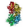
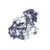
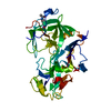
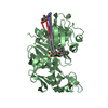
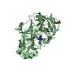
| Entry | Database: PDB / ID: 2v10 | ||||||
|---|---|---|---|---|---|---|---|
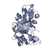
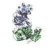
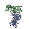
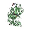
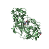
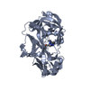
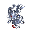
| Title | Crystal Structure of Renin with Inhibitor 9 | ||||||
 Components Components | RENIN | ||||||
 Keywords Keywords | HYDROLASE / GLYCOPROTEIN / INHIBITOR-COMPLEX / ASPARTYL PROTEASE / ZYMOGEN / PROTEASE / POLYMORPHISM / ALTERNATIVE SPLICING / HYDROLASE(ACID PROTEINASE) / CLEAVAGE ON PAIR OF BASIC RESIDUES | ||||||
| Function / homology |  Function and homology information Function and homology informationrenin / mesonephros development / juxtaglomerular apparatus development / renin-angiotensin regulation of aldosterone production / response to cGMP / drinking behavior / response to immobilization stress / regulation of MAPK cascade / amyloid-beta metabolic process / response to cAMP ...renin / mesonephros development / juxtaglomerular apparatus development / renin-angiotensin regulation of aldosterone production / response to cGMP / drinking behavior / response to immobilization stress / regulation of MAPK cascade / amyloid-beta metabolic process / response to cAMP / Metabolism of Angiotensinogen to Angiotensins / angiotensin maturation / insulin-like growth factor receptor binding / cell maturation / hormone-mediated signaling pathway / kidney development / regulation of blood pressure / cellular response to xenobiotic stimulus / male gonad development / apical part of cell / peptidase activity / response to lipopolysaccharide / aspartic-type endopeptidase activity / signaling receptor binding / proteolysis / extracellular space / extracellular region / plasma membrane Similarity search - Function | ||||||
| Biological species |  HOMO SAPIENS (human) HOMO SAPIENS (human) | ||||||
| Method |  X-RAY DIFFRACTION / OTHER / Resolution: 3.1 Å X-RAY DIFFRACTION / OTHER / Resolution: 3.1 Å | ||||||
 Authors Authors | Rahuel, J. / Rasetti, V. / Maibaum, J. / Rueger, H. / Goschke, R. / Cohen, N.C. / Stutz, S. / Cumin, F. / Fuhrer, W. / Wood, J.M. / Grutter, M.G. | ||||||
 Citation Citation | Journal: Chem.Biol. / Year: 2000 Title: Structure-Based Drug Design: The Discovery of Novel Nonpeptide Orally Active Inhibitors of Human Renin Authors: Rahuel, J. / Rasetti, V. / Maibaum, J. / Rueger, H. / Goschke, R. / Cohen, N.C. / Stutz, S. / Cumin, F. / Fuhrer, W. / Wood, J.M. / Grutter, M.G. #1: Journal: J.Struct.Biol. / Year: 1991Title: The Crystal Structures of Recombinant Glycosylated Human Renin Alone and in Complex with a Transition State Analog Inhibitor. Authors: Rahuel, J. / Priestle, J.P. / Grutter, M.G. | ||||||
| History |
| ||||||
| Remark 700 | SHEET THE SHEET STRUCTURE OF THIS MOLECULE IS BIFURCATED. IN ORDER TO REPRESENT THIS FEATURE IN ... SHEET THE SHEET STRUCTURE OF THIS MOLECULE IS BIFURCATED. IN ORDER TO REPRESENT THIS FEATURE IN THE SHEET RECORDS BELOW, TWO SHEETS ARE DEFINED. |
- Structure visualization
| Structure viewer | Molecule: MolmilJmol/JSmol |
|---|
- Downloads & links
-Download
| PDBx/mmCIF format | 2v10.cif.gz | 128.1 KB | Display | PDBx/mmCIF format |
|---|---|---|---|---|
| PDB format | pdb2v10.ent.gz | 95.9 KB | Display | PDB format |
| PDBx/mmJSON format | 2v10.json.gz | Tree view | PDBx/mmJSON format | |
| Others |  Other downloads Other downloads |
-Validation report
| Arichive directory | https://data.pdbj.org/pub/pdb/validation_reports/v1/2v10ftp://data.pdbj.org/pub/pdb/validation_reports/v1/2v10 | HTTPS FTP |
|---|
-Related structure data
| Related structure data |  2v0zC  2v11C  2v12C  2v13C 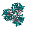 2v16C  1pr7 1pr8 1uhq C: citing same article ( |
|---|---|
| Similar structure data |


-Links
 PDBj
PDBj


- Assembly
| Deposited unit | 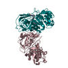
| ||||||||
|---|---|---|---|---|---|---|---|---|---|
| 1 | 
| ||||||||
| 2 | 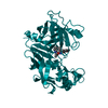
| ||||||||
| Unit cell |
|
-Components
| #1: Protein | Mass: 37267.008 Da / Num. of mol.: 2 Source method: isolated from a genetically manipulated source Source: (gene. exp.) HOMO SAPIENS (human) / Cell line (production host): CHO / Production host:   CRICETULUS GRISEUS (Chinese hamster) / References: UniProt: P00797, renin CRICETULUS GRISEUS (Chinese hamster) / References: UniProt: P00797, renin#2: Chemical |   Mass: 480.680 Da / Num. of mol.: 2 / Source method: obtained synthetically / Formula: C27H48N2O5 Mass: 480.680 Da / Num. of mol.: 2 / Source method: obtained synthetically / Formula: C27H48N2O5Has protein modification | Y | |
|---|
-Experimental details
-Experiment
| Experiment | Method: X-RAY DIFFRACTION |
|---|
- Sample preparation
| Crystal | Density Matthews: 3.27 Å3/Da / Density % sol: 62.38 % |
|---|---|
| Crystal grow | pH: 4.5 / Details: pH 4.50 |
-Data collection
| Diffraction | Mean temperature: 100 K |
|---|---|
| Diffraction source | Source: ROTATING ANODE / Type: ENRAF-NONIUS FR571 / Wavelength: 1.5418 |
| Detector | Type: DELFT INSTRUMENTS / Detector: AREA DETECTOR / Date: Jan 1, 1991 |
| Radiation | Protocol: SINGLE WAVELENGTH / Monochromatic (M) / Laue (L): M / Scattering type: x-ray |
| Radiation wavelength | Wavelength: 1.5418 Å / Relative weight: 1 |
| Reflection | Resolution: 3.1→10 Å / Num. obs: 17734 / % possible obs: 99 % / Observed criterion σ(I): 0 / Redundancy: 3.35 % / Rmerge(I) obs: 0.12 |
- Processing
| Software |
| ||||||||||||
|---|---|---|---|---|---|---|---|---|---|---|---|---|---|
| Refinement | Method to determine structure: OTHER / Resolution: 3.1→10 Å / Cross valid method: THROUGHOUT / σ(F): 0 Details: B-FACTORS WERE NOT REFINED AND A CONSTANT VALUE IS ASSIGNED IN THE COORDINATE-FILE
| ||||||||||||
| Refinement step | Cycle: LAST / Resolution: 3.1→10 Å
|
