 Movie
Movie Controller


+ Open data

- Basic information
| Entry | Database: PDB / ID: 3vyd | ||||||
|---|---|---|---|---|---|---|---|
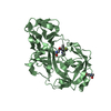
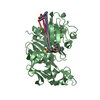
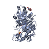
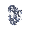
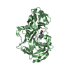
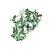
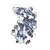
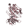
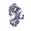
| Title | Human renin in complex with inhibitor 6 | ||||||
 Components Components | Renin | ||||||
 Keywords Keywords | HYDROLASE/HYDROLASE INHIBITOR / Aspartyl protease / RAS / hypertension / inhibitor / HYDROLASE-HYDROLASE INHIBITOR complex | ||||||
| Function / homology |  Function and homology information Function and homology informationrenin / mesonephros development / juxtaglomerular apparatus development / renin-angiotensin regulation of aldosterone production / response to cGMP / drinking behavior / response to immobilization stress / regulation of MAPK cascade / amyloid-beta metabolic process / response to cAMP ...renin / mesonephros development / juxtaglomerular apparatus development / renin-angiotensin regulation of aldosterone production / response to cGMP / drinking behavior / response to immobilization stress / regulation of MAPK cascade / amyloid-beta metabolic process / response to cAMP / Metabolism of Angiotensinogen to Angiotensins / angiotensin maturation / insulin-like growth factor receptor binding / cell maturation / hormone-mediated signaling pathway / kidney development / male gonad development / regulation of blood pressure / cellular response to xenobiotic stimulus / apical part of cell / peptidase activity / response to lipopolysaccharide / aspartic-type endopeptidase activity / signaling receptor binding / proteolysis / : / extracellular region / plasma membrane Similarity search - Function | ||||||
| Biological species |  Homo sapiens (human) Homo sapiens (human) | ||||||
| Method |  X-RAY DIFFRACTION / MOLECULAR REPLACEMENT / Resolution: 2.81 Å X-RAY DIFFRACTION / MOLECULAR REPLACEMENT / Resolution: 2.81 Å | ||||||
 Authors Authors | Takahashi, M. / Hanzawa, H. | ||||||
 Citation Citation | Journal: Bioorg.Med.Chem.Lett. / Year: 2012 Title: Design and discovery of new (3S,5R)-5-[4-(2-chlorophenyl)-2,2-dimethyl-5-oxopiperazin-1-yl]piperidine-3-carboxamides as potent renin inhibitors Authors: Mori, Y. / Ogawa, Y. / Mochizuki, A. / Nakamura, Y. / Sugita, C. / Miyazaki, S. / Tamaki, K. / Matsui, Y. / Takahashi, M. / Nagayama, T. / Nagai, Y. / Inoue, S. / Nishi, T. | ||||||
| History |
|
- Structure visualization
| Structure viewer | Molecule: MolmilJmol/JSmol |
|---|
- Downloads & links
-Download
| PDBx/mmCIF format | 3vyd.cif.gz | 146.6 KB | Display | PDBx/mmCIF format |
|---|---|---|---|---|
| PDB format | pdb3vyd.ent.gz | 116.5 KB | Display | PDB format |
| PDBx/mmJSON format | 3vyd.json.gz | Tree view | PDBx/mmJSON format | |
| Others |  Other downloads Other downloads |
-Validation report
| Arichive directory | https://data.pdbj.org/pub/pdb/validation_reports/vy/3vydftp://data.pdbj.org/pub/pdb/validation_reports/vy/3vyd | HTTPS FTP |
|---|
-Related structure data
| Related structure data |  3vyeC 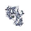 3vyfC 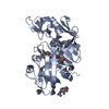 3vswS C: citing same article ( S: Starting model for refinement |
|---|---|
| Similar structure data |


-Links
 PDBj
PDBj


- Assembly
| Deposited unit | 
| ||||||||
|---|---|---|---|---|---|---|---|---|---|
| 1 | 
| ||||||||
| 2 | 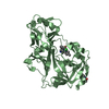
| ||||||||
| 3 | 
| ||||||||
| Unit cell |
|
-Components
| #1: Protein | Mass: 37267.008 Da / Num. of mol.: 2 Source method: isolated from a genetically manipulated source Source: (gene. exp.) Homo sapiens (human) / Gene: REN / Plasmid: pcDNA3.1 / Cell line (production host): 293F / Production host: Homo sapiens (human) / References: UniProt: P00797, renin#2: Sugar |   Type: D-saccharide, beta linking / Mass: 221.208 Da / Num. of mol.: 2 Type: D-saccharide, beta linking / Mass: 221.208 Da / Num. of mol.: 2Source method: isolated from a genetically manipulated source Formula: C8H15NO6 #3: Chemical |   Mass: 449.029 Da / Num. of mol.: 2 / Source method: obtained synthetically / Formula: C24H37ClN4O2 Mass: 449.029 Da / Num. of mol.: 2 / Source method: obtained synthetically / Formula: C24H37ClN4O2#4: Water | ChemComp-HOH / |  Mass: 18.015 Da / Num. of mol.: 169 / Source method: isolated from a natural source / Formula: H2O Mass: 18.015 Da / Num. of mol.: 169 / Source method: isolated from a natural source / Formula: H2OHas protein modification | Y | |
|---|
-Experimental details
-Experiment
| Experiment | Method: X-RAY DIFFRACTION / Number of used crystals: 1 |
|---|
- Sample preparation
| Crystal | Density Matthews: 3.14 Å3/Da / Density % sol: 60.78 % |
|---|---|
| Crystal grow | Temperature: 295 K / Method: vapor diffusion, hanging drop Details: 5-12% PEG 3350, 0.6M NaCl, 0.1M citrate pH3.0-4.5, vapor diffusion, hanging drop, temperature 295K, VAPOR DIFFUSION, HANGING DROP PH range: 3.0-4.5 |
-Data collection
| Diffraction | Mean temperature: 100 K |
|---|---|
| Diffraction source | Source: ROTATING ANODE / Type: RIGAKU FR-E SUPERBRIGHT / Wavelength: 1.502 Å |
| Detector | Type: RIGAKU RAXIS VII / Detector: IMAGE PLATE / Date: Jan 31, 2008 |
| Radiation | Protocol: SINGLE WAVELENGTH / Monochromatic (M) / Laue (L): M / Scattering type: x-ray |
| Radiation wavelength | Wavelength: 1.502 Å / Relative weight: 1 |
| Reflection | Resolution: 2.8→20 Å / Num. obs: 21118 / % possible obs: 91 % |
- Processing
| Software |
| |||||||||||||||||||||||||||||||||||||||||||||
|---|---|---|---|---|---|---|---|---|---|---|---|---|---|---|---|---|---|---|---|---|---|---|---|---|---|---|---|---|---|---|---|---|---|---|---|---|---|---|---|---|---|---|---|---|---|---|
| Refinement | Method to determine structure: MOLECULAR REPLACEMENT Starting model: 3VSW Resolution: 2.81→19.75 Å / Cor.coef. Fo:Fc: 0.94 / Cor.coef. Fo:Fc free: 0.895 / Occupancy max: 1 / Occupancy min: 1 / SU B: 14.886 / SU ML: 0.288 / Cross valid method: THROUGHOUT / σ(F): 0 / ESU R Free: 0.454 / Stereochemistry target values: MAXIMUM LIKELIHOOD Details: HYDROGENS HAVE BEEN USED IF PRESENT IN THE INPUT U VALUES: REFINED INDIVIDUALLY
| |||||||||||||||||||||||||||||||||||||||||||||
| Solvent computation | Ion probe radii: 0.8 Å / Shrinkage radii: 0.8 Å / VDW probe radii: 1.2 Å / Solvent model: BABINET MODEL WITH MASK | |||||||||||||||||||||||||||||||||||||||||||||
| Displacement parameters | Biso max: 118.58 Å2 / Biso mean: 57.8888 Å2 / Biso min: 7.58 Å2 | |||||||||||||||||||||||||||||||||||||||||||||
| Refinement step | Cycle: LAST / Resolution: 2.81→19.75 Å
| |||||||||||||||||||||||||||||||||||||||||||||
| Refine LS restraints |
| |||||||||||||||||||||||||||||||||||||||||||||
| LS refinement shell | Resolution: 2.806→2.877 Å / Total num. of bins used: 20
|
