 Movie
Movie Controller

[English] 日本語
 Yorodumi
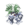
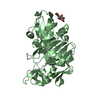
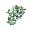
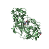
- PDB-5ux4: Crystal Structure of Rat Cathepsin D with (5S)-3-(5,6-dihydro-2H-... -
+ Open data

- Basic information
| Entry | Database: PDB / ID: 5ux4 | |||||||||
|---|---|---|---|---|---|---|---|---|---|---|
| Title | Crystal Structure of Rat Cathepsin D with (5S)-3-(5,6-dihydro-2H-pyran-3-yl)-1-fluoro- 7-(2-fluoropyridin-3-yl)spiro[chromeno[2,3- c]pyridine-5,4'-[1,3]oxazol]-2'-amine | |||||||||
 Components Components | Cathepsin D | |||||||||
 Keywords Keywords | HYDROLASE / Cathepsin D / Beta Amyloid Cleaving Enzyme / BACE1 | |||||||||
| Function / homology |  Function and homology information Function and homology informationCollagen degradation / cathepsin D / aspartic-type peptidase activity / synaptic vesicle endosomal processing / Metabolism of Angiotensinogen to Angiotensins / Insulin receptor recycling / vesicle-mediated transport in synapse / MHC class II antigen presentation / insulin catabolic process / presynaptic endosome ...Collagen degradation / cathepsin D / aspartic-type peptidase activity / synaptic vesicle endosomal processing / Metabolism of Angiotensinogen to Angiotensins / Insulin receptor recycling / vesicle-mediated transport in synapse / MHC class II antigen presentation / insulin catabolic process / presynaptic endosome / regulation of establishment of protein localization / lipoprotein catabolic process / Neutrophil degranulation / insulin receptor recycling / autophagosome assembly / peptide binding / protein catabolic process / endosome lumen / response to nutrient levels / GABA-ergic synapse / melanosome / peptidase activity / endopeptidase activity / aspartic-type endopeptidase activity / lysosome / endosome membrane / positive regulation of apoptotic process / membrane raft / lysosomal membrane / hydrolase activity / proteolysis / : Similarity search - Function | |||||||||
| Biological species |  | |||||||||
| Method |  X-RAY DIFFRACTION / SYNCHROTRON / MOLECULAR REPLACEMENT / molecular replacement / Resolution: 2.805 Å X-RAY DIFFRACTION / SYNCHROTRON / MOLECULAR REPLACEMENT / molecular replacement / Resolution: 2.805 Å | |||||||||
 Authors Authors | Sickmier, A. | |||||||||
 Citation Citation | Journal: Medchemcomm / Year: 2017 Title: Development of 2-aminooxazoline 3-azaxanthene beta-amyloid cleaving enzyme (BACE) inhibitors with improved selectivity against Cathepsin D. Authors: Low, J.D. / Bartberger, M.D. / Chen, K. / Cheng, Y. / Fielden, M.R. / Gore, V. / Hickman, D. / Liu, Q. / Allen Sickmier, E. / Vargas, H.M. / Werner, J. / White, R.D. / Whittington, D.A. / ...Authors: Low, J.D. / Bartberger, M.D. / Chen, K. / Cheng, Y. / Fielden, M.R. / Gore, V. / Hickman, D. / Liu, Q. / Allen Sickmier, E. / Vargas, H.M. / Werner, J. / White, R.D. / Whittington, D.A. / Wood, S. / Minatti, A.E. | |||||||||
| History |
|
- Structure visualization
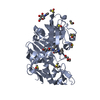
| Structure viewer | Molecule: MolmilJmol/JSmol |
|---|
- Downloads & links
-Download
| PDBx/mmCIF format | 5ux4.cif.gz | 151.4 KB | Display | PDBx/mmCIF format |
|---|---|---|---|---|
| PDB format | pdb5ux4.ent.gz | 115.5 KB | Display | PDB format |
| PDBx/mmJSON format | 5ux4.json.gz | Tree view | PDBx/mmJSON format | |
| Others |  Other downloads Other downloads |
-Validation report
| Arichive directory | https://data.pdbj.org/pub/pdb/validation_reports/ux/5ux4ftp://data.pdbj.org/pub/pdb/validation_reports/ux/5ux4 | HTTPS FTP |
|---|
-Related structure data
| Related structure data | 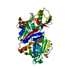 5uyuC 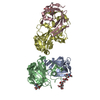 1lyaS S: Starting model for refinement C: citing same article ( |
|---|---|
| Similar structure data |


-Links
 PDBj
PDBj


- Assembly
| Deposited unit | 
| ||||||||
|---|---|---|---|---|---|---|---|---|---|
| 1 | 
| ||||||||
| 2 | 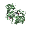
| ||||||||
| Unit cell |
|
-Components
| #1: Protein | Mass: 46825.523 Da / Num. of mol.: 2 Source method: isolated from a genetically manipulated source Source: (gene. exp.)  Cricetulus griseus (Chinese hamster) / References: UniProt: P24268, cathepsin D Cricetulus griseus (Chinese hamster) / References: UniProt: P24268, cathepsin D#2: Polysaccharide | 2-acetamido-2-deoxy-beta-D-glucopyranose-(1-4)-2-acetamido-2-deoxy-beta-D-glucopyranose | Source method: isolated from a genetically manipulated source #3: Sugar |   Type: D-saccharide, beta linking / Mass: 221.208 Da / Num. of mol.: 3 Type: D-saccharide, beta linking / Mass: 221.208 Da / Num. of mol.: 3Source method: isolated from a genetically manipulated source Formula: C8H15NO6 #4: Chemical | 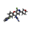  Mass: 448.422 Da / Num. of mol.: 2 / Source method: obtained synthetically / Formula: C24H18F2N4O3 Mass: 448.422 Da / Num. of mol.: 2 / Source method: obtained synthetically / Formula: C24H18F2N4O3#5: Water | ChemComp-HOH / |  Mass: 18.015 Da / Num. of mol.: 25 / Source method: isolated from a natural source / Formula: H2O Mass: 18.015 Da / Num. of mol.: 25 / Source method: isolated from a natural source / Formula: H2OHas protein modification | Y | |
|---|
-Experimental details
-Experiment
| Experiment | Method: X-RAY DIFFRACTION / Number of used crystals: 1 |
|---|
- Sample preparation
| Crystal | Density Matthews: 2.37 Å3/Da / Density % sol: 48.01 % |
|---|---|
| Crystal grow | Temperature: 291 K / Method: vapor diffusion, hanging drop Details: 20% PEG 3350, 2% glycerol, 200 mM Lithium chloride cryo 20% glycerol, 20% PEG 3350, 200 mM lithium chloride |
-Data collection
| Diffraction | Mean temperature: 180 K | ||||||||||||||||||||||||||||||||||||||||||||||||||||||||||||||||||
|---|---|---|---|---|---|---|---|---|---|---|---|---|---|---|---|---|---|---|---|---|---|---|---|---|---|---|---|---|---|---|---|---|---|---|---|---|---|---|---|---|---|---|---|---|---|---|---|---|---|---|---|---|---|---|---|---|---|---|---|---|---|---|---|---|---|---|---|
| Diffraction source | Source: SYNCHROTRON / Site: ALS  / Beamline: 5.0.3 / Wavelength: 1 Å / Beamline: 5.0.3 / Wavelength: 1 Å | ||||||||||||||||||||||||||||||||||||||||||||||||||||||||||||||||||
| Detector | Type: MARMOSAIC 300 mm CCD / Detector: CCD / Date: Nov 8, 2011 | ||||||||||||||||||||||||||||||||||||||||||||||||||||||||||||||||||
| Radiation | Protocol: SINGLE WAVELENGTH / Monochromatic (M) / Laue (L): M / Scattering type: x-ray | ||||||||||||||||||||||||||||||||||||||||||||||||||||||||||||||||||
| Radiation wavelength | Wavelength: 1 Å / Relative weight: 1 | ||||||||||||||||||||||||||||||||||||||||||||||||||||||||||||||||||
| Reflection | Resolution: 2.8→30 Å / Num. obs: 21453 / % possible obs: 99.4 % / Redundancy: 3.6 % / Biso Wilson estimate: 47.07 Å2 / Rmerge(I) obs: 0.095 / Χ2: 1.014 / Net I/σ(I): 9.4 | ||||||||||||||||||||||||||||||||||||||||||||||||||||||||||||||||||
| Reflection shell |
|
-Phasing
| Phasing | Method: molecular replacement |
|---|
- Processing
| Software |
| |||||||||||||||||||||||||||||||||||||||||||||||||||||||||||||||
|---|---|---|---|---|---|---|---|---|---|---|---|---|---|---|---|---|---|---|---|---|---|---|---|---|---|---|---|---|---|---|---|---|---|---|---|---|---|---|---|---|---|---|---|---|---|---|---|---|---|---|---|---|---|---|---|---|---|---|---|---|---|---|---|---|
| Refinement | Method to determine structure: MOLECULAR REPLACEMENT Starting model: 1LYA Resolution: 2.805→29.675 Å / SU ML: 0.24 / Cross valid method: FREE R-VALUE / σ(F): 1.37 / Phase error: 25.06
| |||||||||||||||||||||||||||||||||||||||||||||||||||||||||||||||
| Solvent computation | Shrinkage radii: 0.9 Å / VDW probe radii: 1.11 Å | |||||||||||||||||||||||||||||||||||||||||||||||||||||||||||||||
| Displacement parameters | Biso max: 116.11 Å2 / Biso mean: 46.7496 Å2 / Biso min: 14.84 Å2 | |||||||||||||||||||||||||||||||||||||||||||||||||||||||||||||||
| Refinement step | Cycle: final / Resolution: 2.805→29.675 Å
| |||||||||||||||||||||||||||||||||||||||||||||||||||||||||||||||
| Refine LS restraints |
| |||||||||||||||||||||||||||||||||||||||||||||||||||||||||||||||
| LS refinement shell | Refine-ID: X-RAY DIFFRACTION / Rfactor Rfree error: 0 / Total num. of bins used: 8
|
