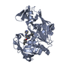
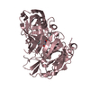
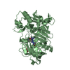
Entry Database : PDB / ID : 5lc0Title Crystal structure of Zika virus NS2B-NS3 protease in complex with a boronate inhibitor NS2B-NS3 protease,NS2B-NS3 protease Keywords / / / / Function / homology Function Domain/homology Component
/ / / / / / / / / / / / / / / / / / / / / / / / / / / / / / / / / / / / / / / / / / / / / / / / / / / / / / / / / / / / / / / / / / / / / / / / / / / / / / / / / / / / / / / / / / / / / / / / / / / / / / / / / / / / / Biological species Method / / / Resolution : 2.7 Å Authors Lei, J. / Hansen, G. / Zhang, L.L. / Hilgenfeld, R. Funding support Organization Grant number Country German Center for Infection Research (DZIF) #TTU01.911_0
Journal : Science / Year : 2016Title : Crystal structure of Zika virus NS2B-NS3 protease in complex with a boronate inhibitor.Authors : Lei, J. / Hansen, G. / Nitsche, C. / Klein, C.D. / Zhang, L. / Hilgenfeld, R. History Deposition Jun 18, 2016 Deposition site / Processing site Revision 1.0 Jul 6, 2016 Provider / Type Revision 1.1 Jul 13, 2016 Group Revision 1.2 Jul 20, 2016 Group Revision 1.3 Aug 10, 2016 Group Revision 1.4 Jan 10, 2024 Group / Database references / Refinement descriptionCategory chem_comp_atom / chem_comp_bond ... chem_comp_atom / chem_comp_bond / database_2 / pdbx_initial_refinement_model Item / _database_2.pdbx_database_accessionRevision 1.5 Oct 23, 2024 Group / Category / pdbx_modification_feature
Show all Show less
 Movie
Movie Controller

 Yorodumi
 Open data
 Basic information
 Components
 Keywords
 Function and homology information
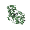
 Zika virus
 X-RAY DIFFRACTION /
  Authors
 Germany, 1items
  Citation
 Structure visualization
 Downloads & links
 Other downloads
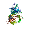


 PDBj
PDBj

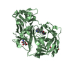
 Assembly
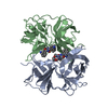

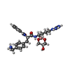
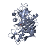
 Mass: 510.394 Da / Num. of mol.: 2 / Source method: obtained synthetically / Formula: C25H35BN6O5
 Mass: 18.015 Da / Num. of mol.: 15 / Source method: isolated from a natural source / Formula: H2O
 Sample preparation
 Processing