 Movie
Movie Controller


+ Open data

- Basic information
| Entry | Database: PDB / ID: 1lyw | ||||||
|---|---|---|---|---|---|---|---|
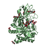
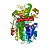
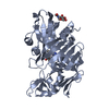
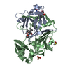
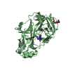
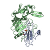
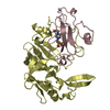
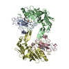
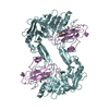
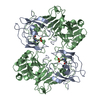
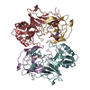
| Title | CATHEPSIN D AT PH 7.5 | ||||||
 Components Components | (CATHEPSIN D) x 2 | ||||||
 Keywords Keywords | ASPARTIC PROTEASE / HYDROLASE / GLYCOPROTEIN | ||||||
| Function / homology |  Function and homology information Function and homology informationcathepsin D / aspartic-type peptidase activity / insulin catabolic process / regulation of establishment of protein localization / lipoprotein catabolic process / execution phase of apoptosis / insulin receptor recycling / Collagen degradation / Metabolism of Angiotensinogen to Angiotensins / autophagosome assembly ...cathepsin D / aspartic-type peptidase activity / insulin catabolic process / regulation of establishment of protein localization / lipoprotein catabolic process / execution phase of apoptosis / insulin receptor recycling / Collagen degradation / Metabolism of Angiotensinogen to Angiotensins / autophagosome assembly / Insulin receptor recycling / MHC class II antigen presentation / lysosomal lumen / endosome lumen / specific granule lumen / antigen processing and presentation of exogenous peptide antigen via MHC class II / tertiary granule lumen / melanosome / peptidase activity / extracellular matrix / Estrogen-dependent gene expression / aspartic-type endopeptidase activity / ficolin-1-rich granule lumen / lysosome / endosome membrane / positive regulation of apoptotic process / membrane raft / lysosomal membrane / cysteine-type endopeptidase activity / Neutrophil degranulation / proteolysis / : / extracellular exosome / extracellular region / cytosol Similarity search - Function | ||||||
| Biological species |  Homo sapiens (human) Homo sapiens (human) | ||||||
| Method |  X-RAY DIFFRACTION / MOLECULAR REPLACEMENT / Resolution: 2.5 Å X-RAY DIFFRACTION / MOLECULAR REPLACEMENT / Resolution: 2.5 Å | ||||||
 Authors Authors | Lee, A.Y. / Gulnik, S.V. / Erickson, J.W. | ||||||
 Citation Citation | Journal: Nat.Struct.Biol. / Year: 1998 Title: Conformational switching in an aspartic proteinase. Authors: Lee, A.Y. / Gulnik, S.V. / Erickson, J.W. #1: Journal: Proc.Natl.Acad.Sci.USA / Year: 1993Title: Crystal Structures of Native and Inhibited Forms of Human Cathepsin D: Implications for Lysosomal Targeting and Drug Design Authors: Baldwin, E.T. / Bhat, T.N. / Gulnik, S. / Hosur, M.V. / Sowder II, R.C. / Cachau, R.E. / Collins, J. / Silva, A.M. / Erickson, J.W. #2: Journal: J.Mol.Biol. / Year: 1992Title: Human Liver Cathepsin D. Purification, Crystallization and Preliminary X-Ray Diffraction Analysis of a Lysosomal Enzyme Authors: Gulnik, S. / Baldwin, E.T. / Tarasova, N. / Erickson, J. | ||||||
| History |
|
- Structure visualization
| Structure viewer | Molecule: MolmilJmol/JSmol |
|---|
- Downloads & links
-Download
| PDBx/mmCIF format | 1lyw.cif.gz | 341 KB | Display | PDBx/mmCIF format |
|---|---|---|---|---|
| PDB format | pdb1lyw.ent.gz | 279.8 KB | Display | PDB format |
| PDBx/mmJSON format | 1lyw.json.gz | Tree view | PDBx/mmJSON format | |
| Others |  Other downloads Other downloads |
-Validation report
| Arichive directory | https://data.pdbj.org/pub/pdb/validation_reports/ly/1lywftp://data.pdbj.org/pub/pdb/validation_reports/ly/1lyw | HTTPS FTP |
|---|
-Related structure data
| Related structure data | 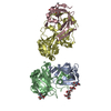 1lyaS S: Starting model for refinement |
|---|---|
| Similar structure data |


-Links
 PDBj
PDBj


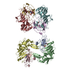
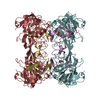
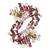
- Assembly


-Components
| #1: Protein | Mass: 10688.941 Da / Num. of mol.: 4 / Source method: isolated from a natural source / Source: (natural) Homo sapiens (human) / Organ: LIVER / References: UniProt: P07339, cathepsin D#2: Protein | Mass: 26270.207 Da / Num. of mol.: 4 / Source method: isolated from a natural source / Source: (natural) Homo sapiens (human) / Organ: LIVER / References: UniProt: P07339, cathepsin D#3: Chemical | ChemComp-EPE /   Mass: 238.305 Da / Num. of mol.: 4 / Source method: obtained synthetically / Formula: C8H18N2O4S / Comment: pH buffer*YM Mass: 238.305 Da / Num. of mol.: 4 / Source method: obtained synthetically / Formula: C8H18N2O4S / Comment: pH buffer*YM#4: Water | ChemComp-HOH / |  Mass: 18.015 Da / Num. of mol.: 558 / Source method: isolated from a natural source / Formula: H2O Mass: 18.015 Da / Num. of mol.: 558 / Source method: isolated from a natural source / Formula: H2OCompound details | CATHEPSIN D IS FOUND PREDOMINANTLY IN A TWO-CHAIN FORM DUE TO A POST-TRANSLATIONAL CLEAVAGE EVENT. ...CATHEPSIN D IS FOUND PREDOMINAN | Has protein modification | Y | |
|---|
-Experimental details
-Experiment
| Experiment | Method: X-RAY DIFFRACTION / Number of used crystals: 1 |
|---|
- Sample preparation
| Crystal | Density Matthews: 1.24 Å3/Da / Density % sol: 65 % / Description: DATA WAS COLLECTED USING 1 DEGREE OSCILLATION | |||||||||||||||||||||||||
|---|---|---|---|---|---|---|---|---|---|---|---|---|---|---|---|---|---|---|---|---|---|---|---|---|---|---|
| Crystal grow | pH: 7.5 / Details: pH 7.5 | |||||||||||||||||||||||||
| Crystal | *PLUS | |||||||||||||||||||||||||
| Crystal grow | *PLUS Temperature: 22 ℃ / Method: vapor diffusion, hanging drop | |||||||||||||||||||||||||
| Components of the solutions | *PLUS
|
-Data collection
| Diffraction | Mean temperature: 190 K |
|---|---|
| Diffraction source | Source: ROTATING ANODE / Type: RIGAKU RUH2R / Wavelength: 1.5418 |
| Detector | Type: MARRESEARCH / Detector: IMAGE PLATE / Date: Jul 1, 1997 / Details: MIRRORS |
| Radiation | Monochromator: NI FILTER / Monochromatic (M) / Laue (L): M / Scattering type: x-ray |
| Radiation wavelength | Wavelength: 1.5418 Å / Relative weight: 1 |
| Reflection | Resolution: 2.5→20 Å / Num. obs: 197513 / % possible obs: 73 % / Observed criterion σ(I): 2 / Redundancy: 3.2 % / Rmerge(I) obs: 0.108 / Rsym value: 0.108 / Net I/σ(I): 6.95 |
| Reflection shell | Resolution: 2.5→2.59 Å / Redundancy: 1.37 % / Rmerge(I) obs: 0.318 / Mean I/σ(I) obs: 2 / Rsym value: 0.318 / % possible all: 41 |
| Reflection | *PLUS Num. obs: 68982 / Num. measured all: 197513 |
- Processing
| Software |
| ||||||||||||||||||||||||||||||||||||||||||||||||||||||||||||
|---|---|---|---|---|---|---|---|---|---|---|---|---|---|---|---|---|---|---|---|---|---|---|---|---|---|---|---|---|---|---|---|---|---|---|---|---|---|---|---|---|---|---|---|---|---|---|---|---|---|---|---|---|---|---|---|---|---|---|---|---|---|
| Refinement | Method to determine structure: MOLECULAR REPLACEMENT Starting model: PDB ENTRY 1LYA Resolution: 2.5→8 Å / Data cutoff high absF: 10000000 / Data cutoff low absF: 0.001 / Cross valid method: THROUGHOUT / σ(F): 2 Details: ALTHOUGH THERE ARE NO NON-CRYSTALLOGRAPHIC SYMMETRIC AXIES. WE DID NOT BOTHER TO LOCATE THEM. THEREFORE, NON-CRYSTALLOGRAPHIC SYMMETRY ARE NOT USED FOR OUR REFINEMENT.
| ||||||||||||||||||||||||||||||||||||||||||||||||||||||||||||
| Refinement step | Cycle: LAST / Resolution: 2.5→8 Å
| ||||||||||||||||||||||||||||||||||||||||||||||||||||||||||||
| Refine LS restraints |
| ||||||||||||||||||||||||||||||||||||||||||||||||||||||||||||
| LS refinement shell | Resolution: 2.5→2.61 Å / Rfactor Rfree error: 0.039 / Total num. of bins used: 8
| ||||||||||||||||||||||||||||||||||||||||||||||||||||||||||||
| Xplor file |
| ||||||||||||||||||||||||||||||||||||||||||||||||||||||||||||
| Software | *PLUS Name: X-PLOR / Version: 3.1 / Classification: refinement | ||||||||||||||||||||||||||||||||||||||||||||||||||||||||||||
| Refine LS restraints | *PLUS
|
