 Movie
Movie Controller

[English] 日本語
 Yorodumi
- PDB-6kxl: Crystal structure of the catalytic domain of Chitiniphilus shinan... -
+ Open data

- Basic information
| Entry | Database: PDB / ID: 6kxl | ||||||||||||||||||||||||
|---|---|---|---|---|---|---|---|---|---|---|---|---|---|---|---|---|---|---|---|---|---|---|---|---|---|
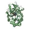
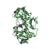
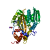
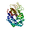
| Title | Crystal structure of the catalytic domain of Chitiniphilus shinanonensis chitinase ChiL (CsChiL) complexed with N,N'-diacetylchitobiose | ||||||||||||||||||||||||
 Components Components | Family 18 chitinase | ||||||||||||||||||||||||
 Keywords Keywords | HYDROLASE / Chitin / Chitinase / Chitinolytic enzyme / Family 18 glycoside hydrolase (GH18) / N-acetylglucosamine (GlcNAc) / Hydrolysis / Transglycosylation / Enzyme-product complex | ||||||||||||||||||||||||
| Function / homology |  Function and homology information Function and homology informationendochitinase activity / chitinase / chitin catabolic process / chitin binding / carbohydrate metabolic process Similarity search - Function | ||||||||||||||||||||||||
| Biological species |  Chitiniphilus shinanonensis (bacteria) Chitiniphilus shinanonensis (bacteria) | ||||||||||||||||||||||||
| Method |  X-RAY DIFFRACTION / SYNCHROTRON / MOLECULAR REPLACEMENT / molecular replacement / Resolution: 1.35 Å X-RAY DIFFRACTION / SYNCHROTRON / MOLECULAR REPLACEMENT / molecular replacement / Resolution: 1.35 Å | ||||||||||||||||||||||||
 Authors Authors | Ueda, M. / Shimosaka, M. / Arai, R. | ||||||||||||||||||||||||
| Funding support |  Japan, 7items Japan, 7items
| ||||||||||||||||||||||||
 Citation Citation | Journal: To be published Title: Crystal structure of CsChiL, a chitinase from Chitiniphilus shinanonensis Authors: Ueda, M. / Sonoda, N. / Shimosaka, M. / Arai, R. #1: Journal: Acta Crystallogr F Struct Biol Commun / Year: 2015 Title: Expression, purification, crystallization and X-ray diffraction analysis of ChiL, a chitinase from Chitiniphilus shinanonensis. Authors: Ueda, M. / Shimosaka, M. / Arai, R. | ||||||||||||||||||||||||
| History |
|
- Structure visualization
| Structure viewer | Molecule: MolmilJmol/JSmol |
|---|
- Downloads & links
-Download
| PDBx/mmCIF format | 6kxl.cif.gz | 363.4 KB | Display | PDBx/mmCIF format |
|---|---|---|---|---|
| PDB format | pdb6kxl.ent.gz | 292.1 KB | Display | PDB format |
| PDBx/mmJSON format | 6kxl.json.gz | Tree view | PDBx/mmJSON format | |
| Others |  Other downloads Other downloads |
-Validation report
| Arichive directory | https://data.pdbj.org/pub/pdb/validation_reports/kx/6kxlftp://data.pdbj.org/pub/pdb/validation_reports/kx/6kxl | HTTPS FTP |
|---|
-Related structure data
| Related structure data |  6kxmC 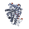 6kxnC  6kstS S: Starting model for refinement C: citing same article ( |
|---|---|
| Similar structure data |


-Links
 PDBj
PDBj- Assembly
| Deposited unit | 
| ||||||||
|---|---|---|---|---|---|---|---|---|---|
| 1 | 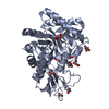
| ||||||||
| 2 | 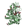
| ||||||||
| Unit cell |
|
-Components
-Protein , 1 types, 2 molecules AB
| #1: Protein | Mass: 41387.938 Da / Num. of mol.: 2 Source method: isolated from a genetically manipulated source Source: (gene. exp.) Chitiniphilus shinanonensis (bacteria) / Strain: SAY3 / Gene: chiL / Plasmid: pCold-ChiL / Production host: |
|---|
-Sugars , 2 types, 4 molecules 
| #2: Polysaccharide | | #8: Sugar |  Type: D-saccharide, beta linking / Mass: 221.208 Da / Num. of mol.: 2 / Source method: obtained synthetically / Formula: C8H15NO6 Type: D-saccharide, beta linking / Mass: 221.208 Da / Num. of mol.: 2 / Source method: obtained synthetically / Formula: C8H15NO6 |
|---|
-Non-polymers , 7 types, 1228 molecules 


| #3: Chemical |  Mass: 59.044 Da / Num. of mol.: 2 / Source method: obtained synthetically / Formula: C2H3O2 Mass: 59.044 Da / Num. of mol.: 2 / Source method: obtained synthetically / Formula: C2H3O2#4: Chemical | ChemComp-MXE / |  Mass: 76.094 Da / Num. of mol.: 1 / Source method: obtained synthetically / Formula: C3H8O2 Mass: 76.094 Da / Num. of mol.: 1 / Source method: obtained synthetically / Formula: C3H8O2#5: Chemical | ChemComp-MLI /  Mass: 102.046 Da / Num. of mol.: 5 / Source method: obtained synthetically / Formula: C3H2O4 Mass: 102.046 Da / Num. of mol.: 5 / Source method: obtained synthetically / Formula: C3H2O4#6: Chemical |  Mass: 150.173 Da / Num. of mol.: 2 / Source method: obtained synthetically / Formula: C6H14O4 Mass: 150.173 Da / Num. of mol.: 2 / Source method: obtained synthetically / Formula: C6H14O4#7: Chemical | ChemComp-EDO /  Mass: 62.068 Da / Num. of mol.: 5 Mass: 62.068 Da / Num. of mol.: 5Source method: isolated from a genetically manipulated source Formula: C2H6O2 #9: Chemical |  Mass: 194.226 Da / Num. of mol.: 3 / Source method: obtained synthetically / Formula: C8H18O5 / Comment: precipitant*YM Mass: 194.226 Da / Num. of mol.: 3 / Source method: obtained synthetically / Formula: C8H18O5 / Comment: precipitant*YM#10: Water | ChemComp-HOH / | Mass: 18.015 Da / Num. of mol.: 1210 / Source method: isolated from a natural source / Formula: H2O |
|---|
-Details
| Has ligand of interest | Y |
|---|---|
| Has protein modification | Y |
-Experimental details
-Experiment
| Experiment | Method: X-RAY DIFFRACTION / Number of used crystals: 1 |
|---|
- Sample preparation
| Crystal | Density Matthews: 2.27 Å3/Da / Density % sol: 45.71 % |
|---|---|
| Crystal grow | Temperature: 293.15 K / Method: vapor diffusion, hanging drop / pH: 7 / Details: 0.2 M sodium malonate, 29% w/v PEG 3350 |
-Data collection
| Diffraction | Mean temperature: 95 K / Serial crystal experiment: N | ||||||||||||||||||||||||||||||||||||||||||||||||||||||||||||||||||||||||||||||||||||||||||||||||||||||||||||||
|---|---|---|---|---|---|---|---|---|---|---|---|---|---|---|---|---|---|---|---|---|---|---|---|---|---|---|---|---|---|---|---|---|---|---|---|---|---|---|---|---|---|---|---|---|---|---|---|---|---|---|---|---|---|---|---|---|---|---|---|---|---|---|---|---|---|---|---|---|---|---|---|---|---|---|---|---|---|---|---|---|---|---|---|---|---|---|---|---|---|---|---|---|---|---|---|---|---|---|---|---|---|---|---|---|---|---|---|---|---|---|---|
| Diffraction source | Source: SYNCHROTRON / Site: Photon Factory / Beamline: AR-NW12A / Wavelength: 1 Å | ||||||||||||||||||||||||||||||||||||||||||||||||||||||||||||||||||||||||||||||||||||||||||||||||||||||||||||||
| Detector | Type: ADSC QUANTUM 210r / Detector: CCD / Date: Dec 13, 2014 / Details: Focusing mirror | ||||||||||||||||||||||||||||||||||||||||||||||||||||||||||||||||||||||||||||||||||||||||||||||||||||||||||||||
| Radiation | Monochromator: Numerical link type Si(111) double crystal / Protocol: SINGLE WAVELENGTH / Monochromatic (M) / Laue (L): M / Scattering type: x-ray | ||||||||||||||||||||||||||||||||||||||||||||||||||||||||||||||||||||||||||||||||||||||||||||||||||||||||||||||
| Radiation wavelength | Wavelength: 1 Å / Relative weight: 1 | ||||||||||||||||||||||||||||||||||||||||||||||||||||||||||||||||||||||||||||||||||||||||||||||||||||||||||||||
| Reflection | Resolution: 1.35→50 Å / Num. obs: 157981 / % possible obs: 95.8 % / Redundancy: 6.7 % / Biso Wilson estimate: 12 Å2 / Rmerge(I) obs: 0.076 / Rpim(I) all: 0.031 / Rrim(I) all: 0.083 / Χ2: 1.015 / Net I/av σ(I): 22.9 / Net I/σ(I): 11.4 | ||||||||||||||||||||||||||||||||||||||||||||||||||||||||||||||||||||||||||||||||||||||||||||||||||||||||||||||
| Reflection shell | Diffraction-ID: 1
|
-Phasing
| Phasing | Method: molecular replacement | |||||||||
|---|---|---|---|---|---|---|---|---|---|---|
| Phasing MR |
|
- Processing
| Software |
| |||||||||||||||||||||||||||||||||||||||||||||||||||||||||||||||||||||||||||||||||||||||||||||||||||||||||||||||||||||||||||||||||||||||||||||||||||
|---|---|---|---|---|---|---|---|---|---|---|---|---|---|---|---|---|---|---|---|---|---|---|---|---|---|---|---|---|---|---|---|---|---|---|---|---|---|---|---|---|---|---|---|---|---|---|---|---|---|---|---|---|---|---|---|---|---|---|---|---|---|---|---|---|---|---|---|---|---|---|---|---|---|---|---|---|---|---|---|---|---|---|---|---|---|---|---|---|---|---|---|---|---|---|---|---|---|---|---|---|---|---|---|---|---|---|---|---|---|---|---|---|---|---|---|---|---|---|---|---|---|---|---|---|---|---|---|---|---|---|---|---|---|---|---|---|---|---|---|---|---|---|---|---|---|---|---|---|
| Refinement | Method to determine structure: MOLECULAR REPLACEMENT Starting model: 6KST Resolution: 1.35→31.21 Å / Cor.coef. Fo:Fc: 0.982 / Cor.coef. Fo:Fc free: 0.975 / Matrix type: sparse / SU B: 1.606 / SU ML: 0.029 / Cross valid method: THROUGHOUT / σ(F): 0 / ESU R: 0.049 / ESU R Free: 0.046 Details: HYDROGENS HAVE BEEN ADDED IN THE RIDING POSITIONS U VALUES : REFINED INDIVIDUALLY
| |||||||||||||||||||||||||||||||||||||||||||||||||||||||||||||||||||||||||||||||||||||||||||||||||||||||||||||||||||||||||||||||||||||||||||||||||||
| Solvent computation | Ion probe radii: 0.8 Å / Shrinkage radii: 0.8 Å / VDW probe radii: 1.2 Å | |||||||||||||||||||||||||||||||||||||||||||||||||||||||||||||||||||||||||||||||||||||||||||||||||||||||||||||||||||||||||||||||||||||||||||||||||||
| Displacement parameters | Biso max: 64.85 Å2 / Biso mean: 15.029 Å2 / Biso min: 8.2 Å2
| |||||||||||||||||||||||||||||||||||||||||||||||||||||||||||||||||||||||||||||||||||||||||||||||||||||||||||||||||||||||||||||||||||||||||||||||||||
| Refinement step | Cycle: final / Resolution: 1.35→31.21 Å
| |||||||||||||||||||||||||||||||||||||||||||||||||||||||||||||||||||||||||||||||||||||||||||||||||||||||||||||||||||||||||||||||||||||||||||||||||||
| Refine LS restraints |
| |||||||||||||||||||||||||||||||||||||||||||||||||||||||||||||||||||||||||||||||||||||||||||||||||||||||||||||||||||||||||||||||||||||||||||||||||||
| LS refinement shell |
|
