Mass: 18.015 Da / Num. of mol.: 321 / Source method: isolated from a natural source / Formula: H2O
-
Experimental details
-
Experiment
Experiment
Method: X-RAY DIFFRACTION / Number of used crystals: 1
-
Sample preparation
Crystal
Density Matthews: 2.88 Å3/Da / Density % sol: 57.23 %
Crystal grow
Temperature: 277 K / Method: vapor diffusion, sitting drop Details: The HODM-heme subunit was crystallised at 3 mg ml-1 in 50 mM KPi, 100 mM KCl, pH 7.5. Initial crystallisation conditions were identified using the JCSG+ matrix screen (Molecular dimensions). ...Details: The HODM-heme subunit was crystallised at 3 mg ml-1 in 50 mM KPi, 100 mM KCl, pH 7.5. Initial crystallisation conditions were identified using the JCSG+ matrix screen (Molecular dimensions). Crystals suitable for diffraction experiments were obtained by sitting drop vapour diffusion at 277 K in 400 nL drops containing equal volumes of protein and a solution containing 30% PEG 2K MME and 0.1 M potassium thiocyanate
Protocol: SINGLE WAVELENGTH / Monochromatic (M) / Laue (L): M / Scattering type: x-ray
Radiation wavelength
Wavelength: 0.917 Å / Relative weight: 1
Reflection
Resolution: 1.65→26.82 Å / Num. obs: 53715 / % possible obs: 99.93 % / Redundancy: 12.1 % / Rmerge(I) obs: 0.061 / Net I/σ(I): 15.4
Reflection shell
Resolution: 1.65→1.7 Å / Redundancy: 12.2 % / Rmerge(I) obs: 0.783 / Mean I/σ(I) obs: 3.8 / % possible all: 100
-
Processing
Software
Name
Version
Classification
REFMAC
5.7.0029
refinement
XDS
datareduction
XDS
datascaling
MLPHARE
phasing
Refinement
Method to determine structure: MIR / Resolution: 1.65→26.82 Å / Cor.coef. Fo:Fc: 0.962 / Cor.coef. Fo:Fc free: 0.948 / SU B: 1.333 / SU ML: 0.046 / Cross valid method: THROUGHOUT / ESU R: 0.076 / ESU R Free: 0.079 / Details: HYDROGENS HAVE BEEN ADDED IN THE RIDING POSITIONS
Rfactor
Num. reflection
% reflection
Selection details
Rfree
0.19359
2872
5.1 %
RANDOM
Rwork
0.16341
-
-
-
obs
0.16493
53715
99.93 %
-
Solvent computation
Ion probe radii: 0.8 Å / Shrinkage radii: 0.8 Å / VDW probe radii: 1.2 Å
 Movie
Movie Controller

 Yorodumi
 Open data
 Basic information
 Components
 Keywords
 Function and homology information
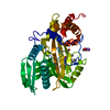
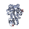
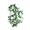
 Pseudomonas mendocina (bacteria)
 X-RAY DIFFRACTION /
  Authors
 United Kingdom, 1items
  Citation
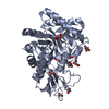
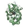
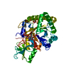
 Structure visualization
 Downloads & links
 Other downloads
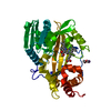


 PDBj
PDBj Assembly
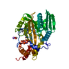

 Mass: 616.487 Da / Num. of mol.: 1 / Source method: obtained synthetically / Formula: C34H32FeN4O4
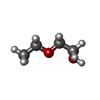
 Mass: 90.121 Da / Num. of mol.: 1 / Source method: obtained synthetically / Formula: C4H10O2
 Mass: 22.990 Da / Num. of mol.: 1 / Source method: obtained synthetically / Formula: Na
 Mass: 18.015 Da / Num. of mol.: 321 / Source method: isolated from a natural source / Formula: H2O
 Sample preparation
 Processing