 Movie
Movie Controller

[English] 日本語
 Yorodumi
- PDB-1htr: CRYSTAL AND MOLECULAR STRUCTURES OF HUMAN PROGASTRICSIN AT 1.62 A... -
+ Open data

- Basic information
| Entry | Database: PDB / ID: 1htr | ||||||
|---|---|---|---|---|---|---|---|
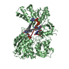
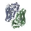
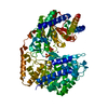
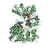
| Title | CRYSTAL AND MOLECULAR STRUCTURES OF HUMAN PROGASTRICSIN AT 1.62 ANGSTROMS RESOLUTION | ||||||
 Components Components |
| ||||||
 Keywords Keywords | ASPARTYL PROTEASE | ||||||
| Function / homology |  Function and homology information Function and homology informationgastricsin / positive regulation of antibacterial peptide production / digestion / aspartic-type endopeptidase activity / proteolysis / : Similarity search - Function | ||||||
| Biological species |  Homo sapiens (human) Homo sapiens (human) | ||||||
| Method |  X-RAY DIFFRACTION / Resolution: 1.62 Å X-RAY DIFFRACTION / Resolution: 1.62 Å | ||||||
 Authors Authors | Moore, S.A. / Sielecki, A.R. / James, M.N.G. | ||||||
 Citation Citation | Journal: J.Mol.Biol. / Year: 1995 Title: Crystal and molecular structures of human progastricsin at 1.62 A resolution. Authors: Moore, S.A. / Sielecki, A.R. / Chernaia, M.M. / Tarasova, N.I. / James, M.N. #1: Journal: Biochim.Biophys.Acta / Year: 1990Title: Isolation, Crystallization and Preliminary X-Ray Diffraction Data of Human Progastricsin Authors: Ivanov, P.K. / Chernaia, M. / Gustchina, A.E. / Pechik, I.V. / Nikonov, S.V. / Tarasova, N.I. | ||||||
| History |
|
- Structure visualization
| Structure viewer | Molecule: MolmilJmol/JSmol |
|---|
- Downloads & links
-Download
| PDBx/mmCIF format | 1htr.cif.gz | 87.8 KB | Display | PDBx/mmCIF format |
|---|---|---|---|---|
| PDB format | pdb1htr.ent.gz | 67.2 KB | Display | PDB format |
| PDBx/mmJSON format | 1htr.json.gz | Tree view | PDBx/mmJSON format | |
| Others |  Other downloads Other downloads |
-Validation report
| Arichive directory | https://data.pdbj.org/pub/pdb/validation_reports/ht/1htrftp://data.pdbj.org/pub/pdb/validation_reports/ht/1htr | HTTPS FTP |
|---|
-Related structure data
| Similar structure data |
|---|


-Links
 PDBj
PDBj
- Assembly
| Deposited unit | 
| ||||||||
|---|---|---|---|---|---|---|---|---|---|
| 1 | 
| ||||||||
| 2 |
| ||||||||
| Unit cell |
| ||||||||
| Atom site foot note | 1: CIS PROLINE - PRO B 23 |
-Components
| #1: Protein/peptide | Mass: 5137.138 Da / Num. of mol.: 1 Source method: isolated from a genetically manipulated source Source: (gene. exp.) Homo sapiens (human) / Tissue: GASTRIC MUCOSA CELL: CHIEF CELLS / Organ: STOMACH / References: UniProt: P20142, gastricsin |
|---|---|
| #2: Protein | Mass: 35464.930 Da / Num. of mol.: 1 Source method: isolated from a genetically manipulated source Source: (gene. exp.) Homo sapiens (human) / Tissue: GASTRIC MUCOSA CELL: CHIEF CELLS / Organ: STOMACH / References: UniProt: P20142, gastricsin |
| #3: Water | ChemComp-HOH /  Mass: 18.015 Da / Num. of mol.: 250 / Source method: isolated from a natural source / Formula: H2O Mass: 18.015 Da / Num. of mol.: 250 / Source method: isolated from a natural source / Formula: H2O |
| Compound details | COMPND THE MOLECULE IS SYNTHESIZED IN THE STOMACH IN THIS INACTIVE ZYMOGEN FORM. AFTER ACTIVATION ...COMPND THE MOLECULE IS SYNTHESIZE |
| Has protein modification | Y |
-Experimental details
-Experiment
| Experiment | Method: X-RAY DIFFRACTION |
|---|
- Sample preparation
| Crystal | Density Matthews: 2.4 Å3/Da / Density % sol: 48.82 % | ||||||||||||||||||||||||||||||
|---|---|---|---|---|---|---|---|---|---|---|---|---|---|---|---|---|---|---|---|---|---|---|---|---|---|---|---|---|---|---|---|
| Crystal grow | *PLUS Method: seeding / PH range low: 6.5 / PH range high: 6 | ||||||||||||||||||||||||||||||
| Components of the solutions | *PLUS
|
-Data collection
| Radiation | Scattering type: x-ray |
|---|---|
| Radiation wavelength | Relative weight: 1 |
| Reflection | Num. obs: 50353 / % possible obs: 96 % / Observed criterion σ(I): 0 |
| Reflection | *PLUS Highest resolution: 1.62 Å / Rmerge(I) obs: 0.059 |
- Processing
| Software |
| ||||||||||||||||||||||||||||||||||||||||||||||||||||||||||||||||||||||||||||||||||||
|---|---|---|---|---|---|---|---|---|---|---|---|---|---|---|---|---|---|---|---|---|---|---|---|---|---|---|---|---|---|---|---|---|---|---|---|---|---|---|---|---|---|---|---|---|---|---|---|---|---|---|---|---|---|---|---|---|---|---|---|---|---|---|---|---|---|---|---|---|---|---|---|---|---|---|---|---|---|---|---|---|---|---|---|---|---|
| Refinement | Resolution: 1.62→20 Å / σ(F): 0 /
| ||||||||||||||||||||||||||||||||||||||||||||||||||||||||||||||||||||||||||||||||||||
| Displacement parameters | Biso mean: 27.75 Å2 | ||||||||||||||||||||||||||||||||||||||||||||||||||||||||||||||||||||||||||||||||||||
| Refinement step | Cycle: LAST / Resolution: 1.62→20 Å
| ||||||||||||||||||||||||||||||||||||||||||||||||||||||||||||||||||||||||||||||||||||
| Refine LS restraints |
| ||||||||||||||||||||||||||||||||||||||||||||||||||||||||||||||||||||||||||||||||||||
| Software | *PLUS Name: PROLSQ/TNT / Classification: refinement | ||||||||||||||||||||||||||||||||||||||||||||||||||||||||||||||||||||||||||||||||||||
| Refinement | *PLUS Num. reflection all: 50353 / Rfactor all: 0.179 | ||||||||||||||||||||||||||||||||||||||||||||||||||||||||||||||||||||||||||||||||||||
| Solvent computation | *PLUS | ||||||||||||||||||||||||||||||||||||||||||||||||||||||||||||||||||||||||||||||||||||
| Displacement parameters | *PLUS |
