 Movie
Movie Controller


+ Open data

- Basic information
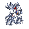
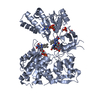
| Entry | Database: PDB / ID: 1j9z | ||||||
|---|---|---|---|---|---|---|---|
| Title | CYPOR-W677G | ||||||
 Components Components | NADPH-Cytochrome P450 reductase | ||||||
 Keywords Keywords | OXIDOREDUCTASE / NADPH-Cytochrome P450 reductase | ||||||
| Function / homology |  Function and homology information Function and homology informationnegative regulation of lipase activity / iron-cytochrome-c reductase activity / positive regulation of growth plate cartilage chondrocyte proliferation / positive regulation of steroid hormone biosynthetic process / nitrate catabolic process / demethylation / organofluorine metabolic process / carnitine metabolic process / cellular response to gonadotropin stimulus / nitric oxide dioxygenase [NAD(P)H] activity ...negative regulation of lipase activity / iron-cytochrome-c reductase activity / positive regulation of growth plate cartilage chondrocyte proliferation / positive regulation of steroid hormone biosynthetic process / nitrate catabolic process / demethylation / organofluorine metabolic process / carnitine metabolic process / cellular response to gonadotropin stimulus / nitric oxide dioxygenase [NAD(P)H] activity / nitric oxide catabolic process / cytochrome-b5 reductase activity, acting on NAD(P)H / positive regulation of chondrocyte differentiation / cellular response to follicle-stimulating hormone stimulus / internal peptidyl-lysine acetylation / NADPH-hemoprotein reductase / NADPH-hemoprotein reductase activity / positive regulation of smoothened signaling pathway / cellular response to peptide hormone stimulus / response to dexamethasone / fatty acid oxidation / nitric oxide biosynthetic process / response to hormone / response to nutrient / electron transport chain / enzyme activator activity / NADP binding / FMN binding / flavin adenine dinucleotide binding / electron transfer activity / oxidoreductase activity / response to xenobiotic stimulus / hydrolase activity / negative regulation of apoptotic process / endoplasmic reticulum membrane / enzyme binding / nucleoplasm / cytosol Similarity search - Function | ||||||
| Biological species |  | ||||||
| Method |  X-RAY DIFFRACTION / FOURIER SYNTHESIS / Resolution: 2.7 Å X-RAY DIFFRACTION / FOURIER SYNTHESIS / Resolution: 2.7 Å | ||||||
 Authors Authors | Hubbard, P.A. / Shen, A.L. / Paschke, R. / Kasper, C.B. / Kim, J.J. | ||||||
 Citation Citation | Journal: J.Biol.Chem. / Year: 2001 Title: NADPH-cytochrome P450 oxidoreductase. Structural basis for hydride and electron transfer. Authors: Hubbard, P.A. / Shen, A.L. / Paschke, R. / Kasper, C.B. / Kim, J.J. | ||||||
| History |
|
- Structure visualization
| Structure viewer | Molecule: MolmilJmol/JSmol |
|---|
- Downloads & links
-Download
| PDBx/mmCIF format | 1j9z.cif.gz | 263.1 KB | Display | PDBx/mmCIF format |
|---|---|---|---|---|
| PDB format | pdb1j9z.ent.gz | 209.7 KB | Display | PDB format |
| PDBx/mmJSON format | 1j9z.json.gz | Tree view | PDBx/mmJSON format | |
| Others |  Other downloads Other downloads |
-Validation report
| Arichive directory | https://data.pdbj.org/pub/pdb/validation_reports/j9/1j9zftp://data.pdbj.org/pub/pdb/validation_reports/j9/1j9z | HTTPS FTP |
|---|
-Related structure data
| Related structure data |  1ja0C  1ja1C 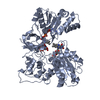 1amoS S: Starting model for refinement C: citing same article ( |
|---|---|
| Similar structure data |


-Links
 PDBj
PDBj

- Assembly
| Deposited unit | 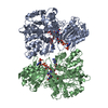
| ||||||||
|---|---|---|---|---|---|---|---|---|---|
| 1 | 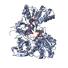
| ||||||||
| 2 | 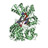
| ||||||||
| Unit cell |
|
-Components
| #1: Protein | Mass: 70551.281 Da / Num. of mol.: 2 / Mutation: W677G Source method: isolated from a genetically manipulated source Source: (gene. exp.)  #2: Chemical |   Mass: 785.550 Da / Num. of mol.: 2 / Source method: obtained synthetically / Formula: C27H33N9O15P2 / Comment: FAD*YM Mass: 785.550 Da / Num. of mol.: 2 / Source method: obtained synthetically / Formula: C27H33N9O15P2 / Comment: FAD*YM#3: Chemical |   Mass: 456.344 Da / Num. of mol.: 2 / Source method: obtained synthetically / Formula: C17H21N4O9P Mass: 456.344 Da / Num. of mol.: 2 / Source method: obtained synthetically / Formula: C17H21N4O9P#4: Chemical |   Mass: 743.405 Da / Num. of mol.: 2 / Source method: obtained synthetically / Formula: C21H28N7O17P3 Mass: 743.405 Da / Num. of mol.: 2 / Source method: obtained synthetically / Formula: C21H28N7O17P3#5: Water | ChemComp-HOH / |  Mass: 18.015 Da / Num. of mol.: 210 / Source method: isolated from a natural source / Formula: H2O Mass: 18.015 Da / Num. of mol.: 210 / Source method: isolated from a natural source / Formula: H2O |
|---|
-Experimental details
-Experiment
| Experiment | Method: X-RAY DIFFRACTION / Number of used crystals: 1 |
|---|
- Sample preparation
| Crystal | Density Matthews: 2.48 Å3/Da / Density % sol: 50.42 % | |||||||||||||||||||||||||||||||||||||||||||||||||||||||||||||||
|---|---|---|---|---|---|---|---|---|---|---|---|---|---|---|---|---|---|---|---|---|---|---|---|---|---|---|---|---|---|---|---|---|---|---|---|---|---|---|---|---|---|---|---|---|---|---|---|---|---|---|---|---|---|---|---|---|---|---|---|---|---|---|---|---|
| Crystal grow | Temperature: 293 K / Method: vapor diffusion, hanging drop / pH: 7 Details: PEG 4000, Hepes, Sodium Acetate, NADP+, pH 7.0, VAPOR DIFFUSION, HANGING DROP, temperature 293K | |||||||||||||||||||||||||||||||||||||||||||||||||||||||||||||||
| Crystal grow | *PLUS | |||||||||||||||||||||||||||||||||||||||||||||||||||||||||||||||
| Components of the solutions | *PLUS
|
-Data collection
| Diffraction | Mean temperature: 100 K |
|---|---|
| Diffraction source | Source: ROTATING ANODE / Type: RIGAKU RU200 / Wavelength: 1.5418 Å |
| Detector | Type: RIGAKU RAXIS IIC / Detector: IMAGE PLATE / Date: Aug 10, 2000 / Details: Mirrors |
| Radiation | Monochromator: Osmic blue confocal mirrors / Protocol: SINGLE WAVELENGTH / Monochromatic (M) / Laue (L): M / Scattering type: x-ray |
| Radiation wavelength | Wavelength: 1.5418 Å / Relative weight: 1 |
| Reflection | Resolution: 2.7→30 Å / Num. all: 38773 / Num. obs: 30991 / % possible obs: 80 % / Observed criterion σ(F): 0 / Observed criterion σ(I): 0 / Redundancy: 11.7 % / Biso Wilson estimate: 31.9 Å2 / Limit h max: 37 / Limit h min: 0 / Limit k max: 42 / Limit k min: 0 / Limit l max: 43 / Limit l min: 0 / Observed criterion F max: 219757.88 / Observed criterion F min: 0.95 / Rmerge(I) obs: 0.081 / Rsym value: 0.081 / Net I/σ(I): 13.9 |
| Reflection shell | Resolution: 2.7→2.75 Å / Redundancy: 1.4 % / Rmerge(I) obs: 0.227 / Mean I/σ(I) obs: 2.4 / Num. unique all: 1099 / Rsym value: 0.227 / % possible all: 57.7 |
| Reflection | *PLUS Num. measured all: 455492 |
| Reflection shell | *PLUS % possible obs: 57.7 % |
- Processing
| Software |
| ||||||||||||||||||||||||||||||||||||||||||||||||||||||||||||||||||||||||||||||||||||||||||
|---|---|---|---|---|---|---|---|---|---|---|---|---|---|---|---|---|---|---|---|---|---|---|---|---|---|---|---|---|---|---|---|---|---|---|---|---|---|---|---|---|---|---|---|---|---|---|---|---|---|---|---|---|---|---|---|---|---|---|---|---|---|---|---|---|---|---|---|---|---|---|---|---|---|---|---|---|---|---|---|---|---|---|---|---|---|---|---|---|---|---|---|
| Refinement | Method to determine structure: FOURIER SYNTHESIS Starting model: PDB Entry 1AMO Resolution: 2.7→30 Å / Rfactor Rfree error: 0.007 / Occupancy max: 1 / Occupancy min: 1 / Isotropic thermal model: Isotropic / Cross valid method: THROUGHOUT / σ(F): 0 / σ(I): 0 / Stereochemistry target values: Engh and Huber
| ||||||||||||||||||||||||||||||||||||||||||||||||||||||||||||||||||||||||||||||||||||||||||
| Solvent computation | Solvent model: CNS bulk solvent model used / Bsol: 10.2752 Å2 / ksol: 0.253768 e/Å3 | ||||||||||||||||||||||||||||||||||||||||||||||||||||||||||||||||||||||||||||||||||||||||||
| Displacement parameters | Biso max: 108.45 Å2 / Biso mean: 44.36 Å2 / Biso min: 6.63 Å2
| ||||||||||||||||||||||||||||||||||||||||||||||||||||||||||||||||||||||||||||||||||||||||||
| Refine analyze |
| ||||||||||||||||||||||||||||||||||||||||||||||||||||||||||||||||||||||||||||||||||||||||||
| Refinement step | Cycle: LAST / Resolution: 2.7→30 Å
| ||||||||||||||||||||||||||||||||||||||||||||||||||||||||||||||||||||||||||||||||||||||||||
| Refine LS restraints |
| ||||||||||||||||||||||||||||||||||||||||||||||||||||||||||||||||||||||||||||||||||||||||||
| LS refinement shell | Refine-ID: X-RAY DIFFRACTION / Total num. of bins used: 8
| ||||||||||||||||||||||||||||||||||||||||||||||||||||||||||||||||||||||||||||||||||||||||||
| Software | *PLUS Name: CNS / Version: 1 / Classification: refinement | ||||||||||||||||||||||||||||||||||||||||||||||||||||||||||||||||||||||||||||||||||||||||||
| Refinement | *PLUS Highest resolution: 2.7 Å / Lowest resolution: 30 Å / σ(F): 0 / % reflection Rfree: 5 % / Rfactor obs: 0.188 / Rfactor Rfree: 0.27 | ||||||||||||||||||||||||||||||||||||||||||||||||||||||||||||||||||||||||||||||||||||||||||
| Solvent computation | *PLUS | ||||||||||||||||||||||||||||||||||||||||||||||||||||||||||||||||||||||||||||||||||||||||||
| Displacement parameters | *PLUS | ||||||||||||||||||||||||||||||||||||||||||||||||||||||||||||||||||||||||||||||||||||||||||
| Refine LS restraints | *PLUS
| ||||||||||||||||||||||||||||||||||||||||||||||||||||||||||||||||||||||||||||||||||||||||||
| LS refinement shell | *PLUS Rfactor Rfree: 0.298 / % reflection Rfree: 3 % / Rfactor Rwork: 0.304 |
