 Movie
Movie Controller

[English] 日本語
 Yorodumi
- PDB-1amo: THREE-DIMENSIONAL STRUCTURE OF NADPH-CYTOCHROME P450 REDUCTASE: P... -
+ Open data

- Basic information
| Entry | Database: PDB / ID: 1amo | ||||||
|---|---|---|---|---|---|---|---|
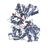
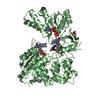
| Title | THREE-DIMENSIONAL STRUCTURE OF NADPH-CYTOCHROME P450 REDUCTASE: PROTOTYPE FOR FMN-AND FAD-CONTAINING ENZYMES | ||||||
 Components Components | NADPH-CYTOCHROME P450 REDUCTASE | ||||||
 Keywords Keywords | OXIDOREDUCTASE / FLAVOPROTEIN / NITRIC OXIDE SYNTHASE | ||||||
| Function / homology |  Function and homology information Function and homology informationiron-cytochrome-c reductase activity / positive regulation of growth plate cartilage chondrocyte proliferation / positive regulation of steroid hormone biosynthetic process / nitrate catabolic process / demethylation / organofluorine metabolic process / carnitine metabolic process / cellular response to gonadotropin stimulus / nitric oxide dioxygenase [NAD(P)H] activity / nitric oxide catabolic process ...iron-cytochrome-c reductase activity / positive regulation of growth plate cartilage chondrocyte proliferation / positive regulation of steroid hormone biosynthetic process / nitrate catabolic process / demethylation / organofluorine metabolic process / carnitine metabolic process / cellular response to gonadotropin stimulus / nitric oxide dioxygenase [NAD(P)H] activity / nitric oxide catabolic process / cytochrome-b5 reductase activity, acting on NAD(P)H / positive regulation of chondrocyte differentiation / cellular response to follicle-stimulating hormone stimulus / NADPH-hemoprotein reductase / NADPH-hemoprotein reductase activity / positive regulation of smoothened signaling pathway / cellular response to peptide hormone stimulus / response to dexamethasone / fatty acid oxidation / nitric oxide biosynthetic process / response to hormone / response to nutrient / electron transport chain / enzyme activator activity / NADP binding / FMN binding / flavin adenine dinucleotide binding / electron transfer activity / oxidoreductase activity / response to xenobiotic stimulus / hydrolase activity / endoplasmic reticulum membrane / negative regulation of apoptotic process / enzyme binding / cytosol Similarity search - Function | ||||||
| Biological species |  | ||||||
| Method |  X-RAY DIFFRACTION / MIR / Resolution: 2.6 Å X-RAY DIFFRACTION / MIR / Resolution: 2.6 Å | ||||||
 Authors Authors | Wang, M. / Roberts, D.L. / Paschke, R. / Shea, T.M. / Masters, B.S.S. / Kim, J.J.P. | ||||||
 Citation Citation | Journal: Proc.Natl.Acad.Sci.USA / Year: 1997 Title: Three-dimensional structure of NADPH-cytochrome P450 reductase: prototype for FMN- and FAD-containing enzymes. Authors: Wang, M. / Roberts, D.L. / Paschke, R. / Shea, T.M. / Masters, B.S. / Kim, J.J. | ||||||
| History |
|
- Structure visualization
| Structure viewer | Molecule: MolmilJmol/JSmol |
|---|
- Downloads & links
-Download
| PDBx/mmCIF format | 1amo.cif.gz | 257.9 KB | Display | PDBx/mmCIF format |
|---|---|---|---|---|
| PDB format | pdb1amo.ent.gz | 205.9 KB | Display | PDB format |
| PDBx/mmJSON format | 1amo.json.gz | Tree view | PDBx/mmJSON format | |
| Others |  Other downloads Other downloads |
-Validation report
| Arichive directory | https://data.pdbj.org/pub/pdb/validation_reports/am/1amoftp://data.pdbj.org/pub/pdb/validation_reports/am/1amo | HTTPS FTP |
|---|
-Related structure data
| Similar structure data |
|---|


-Links
 PDBj
PDBj

- Assembly
| Deposited unit | 
| ||||||||
|---|---|---|---|---|---|---|---|---|---|
| 1 | 
| ||||||||
| 2 | 
| ||||||||
| Unit cell |
| ||||||||
| Noncrystallographic symmetry (NCS) | NCS oper: (Code: given Matrix: (-0.99752, -0.00364, 0.07025), Vector: |
-Components
| #1: Protein | Mass: 69971.641 Da / Num. of mol.: 2 / Fragment: HYDROPHILIC DOMAIN Source method: isolated from a genetically manipulated source Source: (gene. exp.)  #2: Chemical |   Mass: 785.550 Da / Num. of mol.: 2 / Source method: obtained synthetically / Formula: C27H33N9O15P2 / Comment: FAD*YM Mass: 785.550 Da / Num. of mol.: 2 / Source method: obtained synthetically / Formula: C27H33N9O15P2 / Comment: FAD*YM#3: Chemical |   Mass: 456.344 Da / Num. of mol.: 2 / Source method: obtained synthetically / Formula: C17H21N4O9P Mass: 456.344 Da / Num. of mol.: 2 / Source method: obtained synthetically / Formula: C17H21N4O9P#4: Chemical |   Mass: 743.405 Da / Num. of mol.: 2 / Source method: obtained synthetically / Formula: C21H28N7O17P3 Mass: 743.405 Da / Num. of mol.: 2 / Source method: obtained synthetically / Formula: C21H28N7O17P3#5: Water | ChemComp-HOH / |  Mass: 18.015 Da / Num. of mol.: 156 / Source method: isolated from a natural source / Formula: H2O Mass: 18.015 Da / Num. of mol.: 156 / Source method: isolated from a natural source / Formula: H2O |
|---|
-Experimental details
-Experiment
| Experiment | Method: X-RAY DIFFRACTION / Number of used crystals: 1 |
|---|
- Sample preparation
| Crystal | Density Matthews: 2.49 Å3/Da / Density % sol: 49 % | ||||||||||||||||||||||||||||||||||||
|---|---|---|---|---|---|---|---|---|---|---|---|---|---|---|---|---|---|---|---|---|---|---|---|---|---|---|---|---|---|---|---|---|---|---|---|---|---|
| Crystal grow | pH: 7 / Details: pH 7.0 | ||||||||||||||||||||||||||||||||||||
| Crystal grow | *PLUS Temperature: 19 ℃ / pH: 6.5 / Method: vapor diffusionDetails: drop solution was mixed with an equal volume of reservoir solution | ||||||||||||||||||||||||||||||||||||
| Components of the solutions | *PLUS
|
-Data collection
| Diffraction | Mean temperature: 277 K |
|---|---|
| Diffraction source | Source: ROTATING ANODE / Type: RIGAKU RUH2R / Wavelength: 1.5418 |
| Detector | Type: RIGAKU RAXIS IIC / Detector: IMAGE PLATE / Date: Apr 17, 1995 |
| Radiation | Monochromator: GRAPHITE(002) / Monochromatic (M) / Laue (L): M / Scattering type: x-ray |
| Radiation wavelength | Wavelength: 1.5418 Å / Relative weight: 1 |
| Reflection | Resolution: 2.6→30 Å / Num. obs: 41132 / % possible obs: 91.5 % / Observed criterion σ(I): 0 / Redundancy: 4.2 % / Rsym value: 0.07 / Net I/σ(I): 13.5 |
| Reflection shell | Resolution: 2.6→2.76 Å / Redundancy: 1.8 % / Mean I/σ(I) obs: 2.4 / Rsym value: 0.308 / % possible all: 85.8 |
| Reflection | *PLUS Num. measured all: 171499 / Rmerge(I) obs: 0.07 |
| Reflection shell | *PLUS % possible obs: 85.8 % / Rmerge(I) obs: 0.308 |
- Processing
| Software |
| ||||||||||||||||||||||||||||||||||||||||||||||||||||||||||||
|---|---|---|---|---|---|---|---|---|---|---|---|---|---|---|---|---|---|---|---|---|---|---|---|---|---|---|---|---|---|---|---|---|---|---|---|---|---|---|---|---|---|---|---|---|---|---|---|---|---|---|---|---|---|---|---|---|---|---|---|---|---|
| Refinement | Method to determine structure: MIR / Resolution: 2.6→10 Å / Data cutoff high absF: 10000000 / Data cutoff low absF: 0.001 / σ(F): 2
| ||||||||||||||||||||||||||||||||||||||||||||||||||||||||||||
| Displacement parameters | Biso mean: 26.03 Å2 | ||||||||||||||||||||||||||||||||||||||||||||||||||||||||||||
| Refinement step | Cycle: LAST / Resolution: 2.6→10 Å
| ||||||||||||||||||||||||||||||||||||||||||||||||||||||||||||
| Refine LS restraints |
| ||||||||||||||||||||||||||||||||||||||||||||||||||||||||||||
| LS refinement shell | Resolution: 2.6→2.72 Å / Total num. of bins used: 8
| ||||||||||||||||||||||||||||||||||||||||||||||||||||||||||||
| Xplor file |
| ||||||||||||||||||||||||||||||||||||||||||||||||||||||||||||
| Software | *PLUS Name: X-PLOR / Version: 3.8 / Classification: refinement | ||||||||||||||||||||||||||||||||||||||||||||||||||||||||||||
| Refinement | *PLUS | ||||||||||||||||||||||||||||||||||||||||||||||||||||||||||||
| Solvent computation | *PLUS | ||||||||||||||||||||||||||||||||||||||||||||||||||||||||||||
| Displacement parameters | *PLUS | ||||||||||||||||||||||||||||||||||||||||||||||||||||||||||||
| Refine LS restraints | *PLUS
| ||||||||||||||||||||||||||||||||||||||||||||||||||||||||||||
| LS refinement shell | *PLUS Rfactor obs: 0.29 |
