 Movie
Movie Controller

[English] 日本語
 Yorodumi
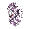
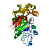
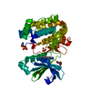
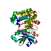
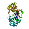
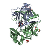
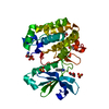
- PDB-2ren: STRUCTURE OF RECOMBINANT HUMAN RENIN, A TARGET FOR CARDIOVASCULAR... -
+ Open data

- Basic information
| Entry | Database: PDB / ID: 2ren | ||||||
|---|---|---|---|---|---|---|---|
| Title | STRUCTURE OF RECOMBINANT HUMAN RENIN, A TARGET FOR CARDIOVASCULAR-ACTIVE DRUGS, AT 2.5 ANGSTROMS RESOLUTION | ||||||
 Components Components | RENIN | ||||||
 Keywords Keywords | HYDROLASE(ACID PROTEINASE) | ||||||
| Function / homology |  Function and homology information Function and homology informationrenin / mesonephros development / juxtaglomerular apparatus development / renin-angiotensin regulation of aldosterone production / response to cGMP / drinking behavior / response to immobilization stress / regulation of MAPK cascade / amyloid-beta metabolic process / response to cAMP ...renin / mesonephros development / juxtaglomerular apparatus development / renin-angiotensin regulation of aldosterone production / response to cGMP / drinking behavior / response to immobilization stress / regulation of MAPK cascade / amyloid-beta metabolic process / response to cAMP / Metabolism of Angiotensinogen to Angiotensins / angiotensin maturation / insulin-like growth factor receptor binding / cell maturation / hormone-mediated signaling pathway / kidney development / male gonad development / regulation of blood pressure / cellular response to xenobiotic stimulus / apical part of cell / peptidase activity / response to lipopolysaccharide / aspartic-type endopeptidase activity / signaling receptor binding / proteolysis / : / extracellular region / plasma membrane Similarity search - Function | ||||||
| Biological species |  Homo sapiens (human) Homo sapiens (human) | ||||||
| Method |  X-RAY DIFFRACTION / Resolution: 2.5 Å X-RAY DIFFRACTION / Resolution: 2.5 Å | ||||||
 Authors Authors | Sielecki, A.R. / James, M.N.G. | ||||||
 Citation Citation | Journal: Science / Year: 1989 Title: Structure of recombinant human renin, a target for cardiovascular-active drugs, at 2.5 A resolution. Authors: Sielecki, A.R. / Hayakawa, K. / Fujinaga, M. / Murphy, M.E. / Fraser, M. / Muir, A.K. / Carilli, C.T. / Lewicki, J.A. / Baxter, J.D. / James, M.N. | ||||||
| History |
| ||||||
| Remark 700 | SHEET THE C-TERMINAL DOMAIN HAS MORE STRANDS THAT ARE NOT FORMALLY HYDROGEN BONDED TO OTHER STRANDS ...SHEET THE C-TERMINAL DOMAIN HAS MORE STRANDS THAT ARE NOT FORMALLY HYDROGEN BONDED TO OTHER STRANDS TO FORM A SHEET. THERE ARE ALSO A FEW IN THE N-TERMINAL DOMAIN. |
- Structure visualization
| Structure viewer | Molecule: MolmilJmol/JSmol |
|---|
- Downloads & links
-Download
| PDBx/mmCIF format | 2ren.cif.gz | 75 KB | Display | PDBx/mmCIF format |
|---|---|---|---|---|
| PDB format | pdb2ren.ent.gz | 55.5 KB | Display | PDB format |
| PDBx/mmJSON format | 2ren.json.gz | Tree view | PDBx/mmJSON format | |
| Others |  Other downloads Other downloads |
-Validation report
| Arichive directory | https://data.pdbj.org/pub/pdb/validation_reports/re/2renftp://data.pdbj.org/pub/pdb/validation_reports/re/2ren | HTTPS FTP |
|---|
-Related structure data
| Similar structure data |
|---|


-Links
 PDBj
PDBj


- Assembly
| Deposited unit | 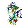
| ||||||||
|---|---|---|---|---|---|---|---|---|---|
| 1 |
| ||||||||
| Unit cell |
| ||||||||
| Atom site foot note | 1: CIS PROLINE - PRO 29 / 2: CIS PROLINE - PRO 308 / 3: CIS PROLINE - PRO 311 4: ATOMS IN THE FOLLOWING RESIDUES HAVE BEEN ASSIGNED A TEMPERATURE FACTOR OF 99.99 INDICATING THAT THE ASSOCIATED ELECTRON DENSITY IS VERY POOR: ARG 82 - GLY 86 SER 213 - THR 214 ALA 248 - ASP 254 |
-Components
| #1: Protein | Mass: 37267.008 Da / Num. of mol.: 1 Source method: isolated from a genetically manipulated source Source: (gene. exp.) Homo sapiens (human) / References: UniProt: P00797, renin |
|---|---|
| #2: Sugar | ChemComp-NAG /   Type: D-saccharide, beta linking / Mass: 221.208 Da / Num. of mol.: 1 Type: D-saccharide, beta linking / Mass: 221.208 Da / Num. of mol.: 1Source method: isolated from a genetically manipulated source Formula: C8H15NO6 |
| Has protein modification | Y |
| Sequence details | THE HUMAN RENIN GENE HAS BEEN SEQUENCED BY TWO GROUPS: 1. HOBART ET AL. (1984) PNAS, V. 81, P. 5026 ...THE HUMAN RENIN GENE HAS BEEN SEQUENCED BY TWO GROUPS: 1. HOBART ET AL. (1984) PNAS, V. 81, P. 5026 2. HARDMAN ET AL. (1984) DNA, V. 3, P. 457 THE EXON5-EXON6 JUNCTION IN 2. HAS A 9 BASE EXON CODING FOR AN ASP-SER-GLU TRIPEPTIDE |
-Experimental details
-Experiment
| Experiment | Method: X-RAY DIFFRACTION |
|---|
- Sample preparation
| Crystal | Density Matthews: 2.53 Å3/Da / Density % sol: 51.39 % | ||||||||||||||||||
|---|---|---|---|---|---|---|---|---|---|---|---|---|---|---|---|---|---|---|---|
| Crystal grow | *PLUS pH: 4.7 / Method: batch method | ||||||||||||||||||
| Components of the solutions | *PLUS
|
-Data collection
| Reflection | *PLUS Highest resolution: 2.5 Å / Lowest resolution: 8 Å / Num. obs: 13343 / Num. measured all: 60512 / Rmerge(I) obs: 0.48 |
|---|
- Processing
| Software |
| ||||||||||||||||||||||||||||||||||||||||||||||||||||||||||||||||||||||||||||||||
|---|---|---|---|---|---|---|---|---|---|---|---|---|---|---|---|---|---|---|---|---|---|---|---|---|---|---|---|---|---|---|---|---|---|---|---|---|---|---|---|---|---|---|---|---|---|---|---|---|---|---|---|---|---|---|---|---|---|---|---|---|---|---|---|---|---|---|---|---|---|---|---|---|---|---|---|---|---|---|---|---|---|
| Refinement | Resolution: 2.5→8 Å / σ(F): 1 Details: ATOMS IN THE FOLLOWING RESIDUES HAVE BEEN ASSIGNED A TEMPERATURE FACTOR OF 99.99 INDICATING THAT THE ASSOCIATED ELECTRON DENSITY IS VERY POOR: ARG 82 - GLY 86 SER 213 - THR 214 ALA 248 - ASP 254
| ||||||||||||||||||||||||||||||||||||||||||||||||||||||||||||||||||||||||||||||||
| Refinement step | Cycle: LAST / Resolution: 2.5→8 Å
| ||||||||||||||||||||||||||||||||||||||||||||||||||||||||||||||||||||||||||||||||
| Refine LS restraints |
| ||||||||||||||||||||||||||||||||||||||||||||||||||||||||||||||||||||||||||||||||
| Refinement | *PLUS Highest resolution: 2.5 Å / Lowest resolution: 8 Å / Num. reflection obs: 13614 / σ(F): 1 / Rfactor obs: 0.217 | ||||||||||||||||||||||||||||||||||||||||||||||||||||||||||||||||||||||||||||||||
| Solvent computation | *PLUS | ||||||||||||||||||||||||||||||||||||||||||||||||||||||||||||||||||||||||||||||||
| Displacement parameters | *PLUS | ||||||||||||||||||||||||||||||||||||||||||||||||||||||||||||||||||||||||||||||||
| Refine LS restraints | *PLUS
|
