 Movie
Movie Controller

[English] 日本語
 Yorodumi
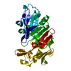
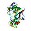
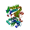
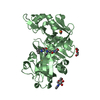
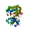
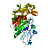
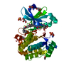
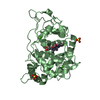
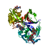
- PDB-1pso: The crystal structure of human pepsin and its complex with pepstatin -
+ Open data

- Basic information
| Entry | Database: PDB / ID: 1pso | ||||||
|---|---|---|---|---|---|---|---|
| Title | The crystal structure of human pepsin and its complex with pepstatin | ||||||
 Components Components |
| ||||||
 Keywords Keywords | HYDROLASE/HYDROLASE INHIBITOR / ACID PROTEINASE / HYDROLASE-HYDROLASE INHIBITOR complex | ||||||
| Function / homology |  Function and homology information Function and homology informationmultivesicular body lumen / pepsin A / Surfactant metabolism / digestion / aspartic-type endopeptidase activity / proteolysis / extracellular exosome Similarity search - Function | ||||||
| Biological species |  Homo sapiens (human) Homo sapiens (human) Streptomyces argenteolus subsp. toyonakensis (bacteria) Streptomyces argenteolus subsp. toyonakensis (bacteria) | ||||||
| Method |  X-RAY DIFFRACTION / Resolution: 2 Å X-RAY DIFFRACTION / Resolution: 2 Å | ||||||
 Authors Authors | Fujinaga, M. / Chernaia, M.M. / Tarasova, N. / Mosimann, S.C. / James, M.N.G. | ||||||
 Citation Citation | Journal: Protein Sci. / Year: 1995 Title: Crystal structure of human pepsin and its complex with pepstatin. Authors: Fujinaga, M. / Chernaia, M.M. / Tarasova, N.I. / Mosimann, S.C. / James, M.N. #1: Journal: J.Mol.Biol. / Year: 1990Title: Molecular and Crystal Structures of Monoclinic Porcine Pepsin Refined at 1.8 Angstroms Resolution Authors: Sielecki, A.R. / Fedorov, A.A. / Boodhoo, A. / Andreeva, N.S. / James, M.N.G. | ||||||
| History |
|
- Structure visualization
| Structure viewer | Molecule: MolmilJmol/JSmol |
|---|
- Downloads & links
-Download
| PDBx/mmCIF format | 1pso.cif.gz | 75.2 KB | Display | PDBx/mmCIF format |
|---|---|---|---|---|
| PDB format | pdb1pso.ent.gz | 53.8 KB | Display | PDB format |
| PDBx/mmJSON format | 1pso.json.gz | Tree view | PDBx/mmJSON format | |
| Others |  Other downloads Other downloads |
-Validation report
| Arichive directory | https://data.pdbj.org/pub/pdb/validation_reports/ps/1psoftp://data.pdbj.org/pub/pdb/validation_reports/ps/1pso | HTTPS FTP |
|---|
-Related structure data


-Links
 PDBj
PDBj
- Assembly
| Deposited unit | 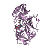
| ||||||||
|---|---|---|---|---|---|---|---|---|---|
| 1 |
| ||||||||
| Unit cell |
| ||||||||
| Atom site foot note | 1: CIS PROLINE - PRO E 23 |
-Components
| #1: Protein | Mass: 34631.887 Da / Num. of mol.: 1 Source method: isolated from a genetically manipulated source Source: (gene. exp.) Homo sapiens (human) / References: UniProt: P00790, UniProt: P0DJD7*PLUS, pepsin A |
|---|---|
| #2: Protein/peptide | 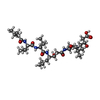  Type: Oligopeptide / Class: Enzyme inhibitor / Mass: 685.891 Da / Num. of mol.: 1 Type: Oligopeptide / Class: Enzyme inhibitor / Mass: 685.891 Da / Num. of mol.: 1Source method: isolated from a genetically manipulated source Source: (gene. exp.) Streptomyces argenteolus subsp. toyonakensis (bacteria)References: Pepstatin |
| #3: Water | ChemComp-HOH /  Mass: 18.015 Da / Num. of mol.: 234 / Source method: isolated from a natural source / Formula: H2O Mass: 18.015 Da / Num. of mol.: 234 / Source method: isolated from a natural source / Formula: H2O |
| Has protein modification | Y |
-Experimental details
-Experiment
| Experiment | Method: X-RAY DIFFRACTION / Number of used crystals: 1 |
|---|
- Sample preparation
| Crystal | Density Matthews: 3.24 Å3/Da / Density % sol: 59.7 % | ||||||||||||||||||||
|---|---|---|---|---|---|---|---|---|---|---|---|---|---|---|---|---|---|---|---|---|---|
| Crystal grow | *PLUS Method: vapor diffusion, hanging drop | ||||||||||||||||||||
| Components of the solutions | *PLUS
|
-Data collection
| Diffraction source | Source: ROTATING ANODE / Wavelength: 1.5418 Å |
|---|---|
| Detector | Type: SAN DIEGO MULTIWIRE DETECTION SYSTEM / Detector: AREA DETECTOR / Date: Jun 17, 1994 |
| Radiation | Scattering type: x-ray |
| Radiation wavelength | Wavelength: 1.5418 Å / Relative weight: 1 |
| Reflection | Num. obs: 31184 / % possible obs: 97 % / Observed criterion σ(I): 0 |
| Reflection | *PLUS Highest resolution: 1.97 Å / Num. measured all: 166144 / Rmerge(I) obs: 0.054 |
- Processing
| Software |
| ||||||||||||
|---|---|---|---|---|---|---|---|---|---|---|---|---|---|
| Refinement | Resolution: 2→30 Å / σ(F): 0 Details: RESIDUES WITH ZERO OCCUPANCIES AND NEGATIVE B-FACTORS ARE DISORDERED AND THEIR POSITIONS ARE NOT CONSIDERED TO HAVE BEEN DETERMINED.
| ||||||||||||
| Displacement parameters | Biso mean: 23 Å2 | ||||||||||||
| Refinement step | Cycle: LAST / Resolution: 2→30 Å
| ||||||||||||
| Refine LS restraints |
| ||||||||||||
| Refine LS restraints | *PLUS
|
