Activation of NOXA and translocation to mitochondria / BH3-only proteins associate with and inactivate anti-apoptotic BCL-2 members / response to dsRNA / positive regulation of extrinsic apoptotic signaling pathway via death domain receptors / positive regulation of protein localization to mitochondrion / positive regulation of glucose metabolic process / positive regulation of oxidative stress-induced neuron intrinsic apoptotic signaling pathway / regulation of mitochondrial membrane permeability / Bcl-2 family protein complex / positive regulation of DNA damage response, signal transduction by p53 class mediator ...Activation of NOXA and translocation to mitochondria / BH3-only proteins associate with and inactivate anti-apoptotic BCL-2 members / response to dsRNA / positive regulation of extrinsic apoptotic signaling pathway via death domain receptors / positive regulation of protein localization to mitochondrion / positive regulation of glucose metabolic process / positive regulation of oxidative stress-induced neuron intrinsic apoptotic signaling pathway / regulation of mitochondrial membrane permeability / Bcl-2 family protein complex / positive regulation of DNA damage response, signal transduction by p53 class mediator / positive regulation of release of cytochrome c from mitochondria / apoptotic mitochondrial changes / T cell homeostasis / negative regulation of mitochondrial membrane potential / intrinsic apoptotic signaling pathway by p53 class mediator / negative regulation of anoikis / BH3 domain binding / intrinsic apoptotic signaling pathway in response to endoplasmic reticulum stress / intrinsic apoptotic signaling pathway in response to DNA damage by p53 class mediator / negative regulation of extrinsic apoptotic signaling pathway in absence of ligand / response to X-ray / response to UV / negative regulation of fibroblast proliferation / cellular response to glucose starvation / proteasomal protein catabolic process / positive regulation of intrinsic apoptotic signaling pathway / extrinsic apoptotic signaling pathway in absence of ligand / response to cytokine / negative regulation of autophagy / intrinsic apoptotic signaling pathway / reactive oxygen species metabolic process / release of cytochrome c from mitochondria / intrinsic apoptotic signaling pathway in response to DNA damage / positive regulation of neuron apoptotic process / regulation of apoptotic process / defense response to virus / cell differentiation / positive regulation of apoptotic process / mitochondrial matrix / protein heterodimerization activity / apoptotic process / DNA damage response / negative regulation of apoptotic process / mitochondrion / nucleoplasm / membrane / nucleus / cytoplasm / cytosol Similarity search - Function
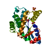
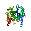
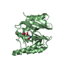
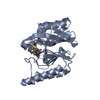
Phorbol-12-myristate-13-acetate-induced protein 1 / Phorbol-12-myristate-13-acetate-induced / Apoptosis regulator, Mcl-1 / Blc2-like / Apoptosis Regulator Bcl-x / Apoptosis regulator, Bcl-2, BH3 motif, conserved site / Apoptosis regulator, Bcl-2 family BH3 motif signature. / Apoptosis regulator, Bcl-2, BH1 motif, conserved site / Apoptosis regulator, Bcl-2 family BH1 motif signature. / Apoptosis regulator, Bcl-2, BH2 motif, conserved site ...Phorbol-12-myristate-13-acetate-induced protein 1 / Phorbol-12-myristate-13-acetate-induced / Apoptosis regulator, Mcl-1 / Blc2-like / Apoptosis Regulator Bcl-x / Apoptosis regulator, Bcl-2, BH3 motif, conserved site / Apoptosis regulator, Bcl-2 family BH3 motif signature. / Apoptosis regulator, Bcl-2, BH1 motif, conserved site / Apoptosis regulator, Bcl-2 family BH1 motif signature. / Apoptosis regulator, Bcl-2, BH2 motif, conserved site / Apoptosis regulator, Bcl-2 family BH2 motif signature. / Bcl-2 family / BCL (B-Cell lymphoma); contains BH1, BH2 regions / Bcl2-like / Bcl-2, Bcl-2 homology region 1-3 / Apoptosis regulator proteins, Bcl-2 family / BCL2-like apoptosis inhibitors family profile. / Bcl-2-like superfamily / Orthogonal Bundle / Mainly Alpha Similarity search - Domain/homology
Induced myeloid leukemia cell differentiation protein Mcl-1 homolog / Phorbol-12-myristate-13-acetate-induced protein 1 Similarity search - Component
Ionic strength: 120 mM / pH: 6.7 / Pressure: ambient / Temperature: 298 K
-
NMR measurement
NMR spectrometer
Type
Manufacturer
Model
Field strength (MHz)
Spectrometer-ID
Bruker Avance
Bruker
AVANCE
500
1
Bruker DRX
Bruker
DRX
600
2
Bruker Avance
Bruker
AVANCE
800
3
-
Processing
NMR software
Name
Version
Developer
Classification
TopSpin
1.3
BrukerBiospin
collection
TopSpin
1.3
BrukerBiospin
processing
XEASY
1.3
Bartelsetal.
peakpicking
XEASY
1.3
Bartelsetal.
chemicalshiftassignment
XEASY
1.3
Bartelsetal.
dataanalysis
CYANA
2.1
Guntert, MumenthalerandWuthrich
structuresolution
X-PLOR NIH
Schwieters, Kuszewski, TjandraandClore
refinement
Refinement
Method: DGSA-distance geometry simulated annealing / Software ordinal: 1 Details: The structures were calculated based on a total of 3355 constraints. 2662 are NOE derived and 184 of these are intermolecular. A total of 325 dihedral angle constraints were used and 184 hydrogen bonds.
NMR constraints
NOE constraints total: 2662 / NOE intraresidue total count: 849 / NOE long range total count: 725 / NOE medium range total count: 545 / NOE sequential total count: 543 / Hydrogen bond constraints total count: 184 / Protein phi angle constraints total count: 175 / Protein psi angle constraints total count: 150
NMR representative
Selection criteria: closest to the average
NMR ensemble
Conformer selection criteria: structures with the least restraint violations Conformers calculated total number: 256 / Conformers submitted total number: 20 / Maximum torsion angle constraint violation: 5 ° / Maximum upper distance constraint violation: 0.2 Å
NMR ensemble rms
Distance rms dev: 0.01 Å / Distance rms dev error: 0.0009 Å
+
About Yorodumi
-
News
-
Feb 9, 2022. New format data for meta-information of EMDB entries
New format data for meta-information of EMDB entries
Version 3 of the EMDB header file is now the official format.
The previous official version 1.9 will be removed from the archive.
In the structure databanks used in Yorodumi, some data are registered as the other names, "COVID-19 virus" and "2019-nCoV". Here are the details of the virus and the list of structure data.
Jan 31, 2019. EMDB accession codes are about to change! (news from PDBe EMDB page)
EMDB accession codes are about to change! (news from PDBe EMDB page)
The allocation of 4 digits for EMDB accession codes will soon come to an end. Whilst these codes will remain in use, new EMDB accession codes will include an additional digit and will expand incrementally as the available range of codes is exhausted. The current 4-digit format prefixed with “EMD-” (i.e. EMD-XXXX) will advance to a 5-digit format (i.e. EMD-XXXXX), and so on. It is currently estimated that the 4-digit codes will be depleted around Spring 2019, at which point the 5-digit format will come into force.
The EM Navigator/Yorodumi systems omit the EMD- prefix.
Related info.:Q: What is EMD? / ID/Accession-code notation in Yorodumi/EM Navigator
Yorodumi is a browser for structure data from EMDB, PDB, SASBDB, etc.
This page is also the successor to EM Navigator detail page, and also detail information page/front-end page for Omokage search.
The word "yorodu" (or yorozu) is an old Japanese word meaning "ten thousand". "mi" (miru) is to see.
Related info.:EMDB / PDB / SASBDB / Comparison of 3 databanks / Yorodumi Search / Aug 31, 2016. New EM Navigator & Yorodumi / Yorodumi Papers / Jmol/JSmol / Function and homology information / Changes in new EM Navigator and Yorodumi
 Movie
Movie Controller


 Open data
 Basic information
 Components
 Keywords
 Function and homology information
 Authors
 Citation
 Structure visualization
 Downloads & links
 Other downloads


 PDBj
PDBj
 Assembly
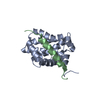

 Sample preparation
 Processing
 CYANA
CYANA