 Movie
Movie Controller


+ Open data

- Basic information
| Entry | Database: PDB / ID: 2r0k | ||||||
|---|---|---|---|---|---|---|---|
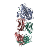
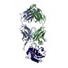
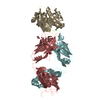
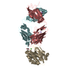
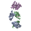
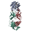
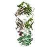
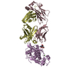
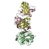
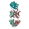
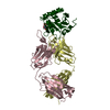
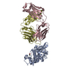
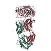
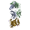
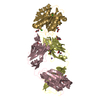
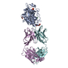
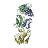
| Title | Protease domain of HGFA with inhibitor Fab58 | ||||||
 Components Components |
| ||||||
 Keywords Keywords | HYDROLASE / IMMUNE SYSTEM / serine protease / antibody / inhibitor / EGF-like domain / Glycoprotein / Kringle / Secreted / Zymogen | ||||||
| Function / homology |  Function and homology information Function and homology informationMET Receptor Activation / zymogen activation / Hydrolases; Acting on peptide bonds (peptidases); Serine endopeptidases / rough endoplasmic reticulum / serine-type peptidase activity / blood coagulation / serine-type endopeptidase activity / proteolysis / extracellular space / extracellular region / cytosol Similarity search - Function | ||||||
| Biological species |  Homo sapiens (human) Homo sapiens (human) | ||||||
| Method |  X-RAY DIFFRACTION / SYNCHROTRON / MOLECULAR REPLACEMENT / Resolution: 3.51 Å X-RAY DIFFRACTION / SYNCHROTRON / MOLECULAR REPLACEMENT / Resolution: 3.51 Å | ||||||
 Authors Authors | Eigenbrot, C. / Shia, S. | ||||||
 Citation Citation | Journal: Proc.Natl.Acad.Sci.Usa / Year: 2007 Title: Structural insight into distinct mechanisms of protease inhibition by antibodies. Authors: Wu, Y. / Eigenbrot, C. / Liang, W.C. / Stawicki, S. / Shia, S. / Fan, B. / Ganesan, R. / Lipari, M.T. / Kirchhofer, D. | ||||||
| History |
|
- Structure visualization
| Structure viewer | Molecule: MolmilJmol/JSmol |
|---|
- Downloads & links
-Download
| PDBx/mmCIF format | 2r0k.cif.gz | 134.9 KB | Display | PDBx/mmCIF format |
|---|---|---|---|---|
| PDB format | pdb2r0k.ent.gz | 104 KB | Display | PDB format |
| PDBx/mmJSON format | 2r0k.json.gz | Tree view | PDBx/mmJSON format | |
| Others |  Other downloads Other downloads |
-Validation report
| Arichive directory | https://data.pdbj.org/pub/pdb/validation_reports/r0/2r0kftp://data.pdbj.org/pub/pdb/validation_reports/r0/2r0k | HTTPS FTP |
|---|
-Related structure data
| Related structure data |  2r0lC 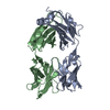 1fvdS  1ybwS S: Starting model for refinement C: citing same article ( |
|---|---|
| Similar structure data |


-Links
 PDBj
PDBj


- Assembly
| Deposited unit | 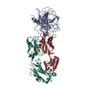
| ||||||||
|---|---|---|---|---|---|---|---|---|---|
| 1 |
| ||||||||
| Unit cell |
|
-Components
| #1: Protein | Mass: 30885.154 Da / Num. of mol.: 1 / Fragment: HGFA protease domain Source method: isolated from a genetically manipulated source Source: (gene. exp.) Homo sapiens (human) / Gene: HGFAC / Production host:   Spodoptera frugiperda (fall armyworm) Spodoptera frugiperda (fall armyworm)References: UniProt: Q04756, Hydrolases; Acting on peptide bonds (peptidases); Serine endopeptidases |
|---|---|
| #2: Antibody | Mass: 23287.793 Da / Num. of mol.: 1 Source method: isolated from a genetically manipulated source Source: (gene. exp.) Homo sapiens (human)Description: The protein was made using a synthetically diversified gene library and selected for tight binding to a specific target on a plastic surface. The gene library used cloned human genes as its basis Production host:  |
| #3: Antibody | Mass: 23688.484 Da / Num. of mol.: 1 Source method: isolated from a genetically manipulated source Source: (gene. exp.) Homo sapiens (human)Description: The protein was made using a synthetically diversified gene library and selected for tight binding to a specific target on a plastic surface. The gene library used cloned human genes as its basis. Production host: |
| Has protein modification | Y |
-Experimental details
-Experiment
| Experiment | Method: X-RAY DIFFRACTION / Number of used crystals: 1 |
|---|
- Sample preparation
| Crystal | Density Matthews: 3.16 Å3/Da / Density % sol: 61.12 % |
|---|---|
| Crystal grow | Method: vapor diffusion, sitting drop / pH: 9.5 Details: 1:1 mixture of protein complex solution and reservoir containing 1.0M K/Na tartrate, CHES pH9.5, 0.2M Lithium Sulfate, VAPOR DIFFUSION, SITTING DROP |
-Data collection
| Diffraction | Mean temperature: 100 K |
|---|---|
| Diffraction source | Source: SYNCHROTRON / Site: ALS  / Beamline: 5.0.2 / Wavelength: 1 Å / Beamline: 5.0.2 / Wavelength: 1 Å |
| Detector | Type: ADSC QUANTUM 315 / Detector: CCD / Date: Apr 5, 2007 |
| Radiation | Monochromator: Si 111 channel / Protocol: SINGLE WAVELENGTH / Monochromatic (M) / Laue (L): M / Scattering type: x-ray |
| Radiation wavelength | Wavelength: 1 Å / Relative weight: 1 |
| Reflection | Resolution: 3.5→50 Å / Num. all: 10827 / Num. obs: 10827 / % possible obs: 88 % / Observed criterion σ(F): 0 / Observed criterion σ(I): -2 / Redundancy: 2.8 % / Biso Wilson estimate: 9 Å2 / Rmerge(I) obs: 0.128 / Net I/σ(I): 6.8 |
| Reflection shell | Resolution: 3.5→3.63 Å / Redundancy: 2.7 % / Rmerge(I) obs: 0.3 / Mean I/σ(I) obs: 2.5 / % possible all: 85.5 |
- Processing
| Software |
| ||||||||||||||||||||||||||||||||||||||||||||||||||||||||||||||||||||||||||||||||||||||||||||||||||||||||||||||||||||||||||||||||||||||||||||||||||||||
|---|---|---|---|---|---|---|---|---|---|---|---|---|---|---|---|---|---|---|---|---|---|---|---|---|---|---|---|---|---|---|---|---|---|---|---|---|---|---|---|---|---|---|---|---|---|---|---|---|---|---|---|---|---|---|---|---|---|---|---|---|---|---|---|---|---|---|---|---|---|---|---|---|---|---|---|---|---|---|---|---|---|---|---|---|---|---|---|---|---|---|---|---|---|---|---|---|---|---|---|---|---|---|---|---|---|---|---|---|---|---|---|---|---|---|---|---|---|---|---|---|---|---|---|---|---|---|---|---|---|---|---|---|---|---|---|---|---|---|---|---|---|---|---|---|---|---|---|---|---|---|---|
| Refinement | Method to determine structure: MOLECULAR REPLACEMENT Starting model: pdb 1YBW, pdb 1FVD Resolution: 3.51→50 Å / Cor.coef. Fo:Fc: 0.83 / Cor.coef. Fo:Fc free: 0.746 / SU B: 81.71 / SU ML: 0.585 / TLS residual ADP flag: LIKELY RESIDUAL / Isotropic thermal model: isotropic / Cross valid method: THROUGHOUT / σ(F): 0 / σ(I): -2 / ESU R Free: 0.813 / Stereochemistry target values: MAXIMUM LIKELIHOOD
| ||||||||||||||||||||||||||||||||||||||||||||||||||||||||||||||||||||||||||||||||||||||||||||||||||||||||||||||||||||||||||||||||||||||||||||||||||||||
| Solvent computation | Ion probe radii: 0.8 Å / Shrinkage radii: 0.8 Å / VDW probe radii: 1.2 Å / Solvent model: MASK | ||||||||||||||||||||||||||||||||||||||||||||||||||||||||||||||||||||||||||||||||||||||||||||||||||||||||||||||||||||||||||||||||||||||||||||||||||||||
| Displacement parameters | Biso mean: 16.411 Å2
| ||||||||||||||||||||||||||||||||||||||||||||||||||||||||||||||||||||||||||||||||||||||||||||||||||||||||||||||||||||||||||||||||||||||||||||||||||||||
| Refinement step | Cycle: LAST / Resolution: 3.51→50 Å
| ||||||||||||||||||||||||||||||||||||||||||||||||||||||||||||||||||||||||||||||||||||||||||||||||||||||||||||||||||||||||||||||||||||||||||||||||||||||
| Refine LS restraints |
| ||||||||||||||||||||||||||||||||||||||||||||||||||||||||||||||||||||||||||||||||||||||||||||||||||||||||||||||||||||||||||||||||||||||||||||||||||||||
| LS refinement shell | Resolution: 3.508→3.599 Å / Total num. of bins used: 20
| ||||||||||||||||||||||||||||||||||||||||||||||||||||||||||||||||||||||||||||||||||||||||||||||||||||||||||||||||||||||||||||||||||||||||||||||||||||||
| Refinement TLS params. | Method: refined / Refine-ID: X-RAY DIFFRACTION
| ||||||||||||||||||||||||||||||||||||||||||||||||||||||||||||||||||||||||||||||||||||||||||||||||||||||||||||||||||||||||||||||||||||||||||||||||||||||
| Refinement TLS group |
|
