 Movie
Movie Controller

[English] 日本語
 Yorodumi
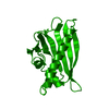
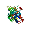
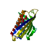
- PDB-2qim: Crystal Structure of Pathogenesis-related Protein LlPR-10.2B from... -
+ Open data

- Basic information
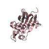
| Entry | Database: PDB / ID: 2qim | ||||||
|---|---|---|---|---|---|---|---|
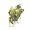
| Title | Crystal Structure of Pathogenesis-related Protein LlPR-10.2B from yellow lupine in complex with Cytokinin | ||||||
 Components Components | PR10.2B | ||||||
 Keywords Keywords | ALLERGEN / trans-zeatin / cytokinin / plant hormones / plant protein / PR-10 protein / pathogenesis-related protein | ||||||
| Function / homology |  Function and homology information Function and homology informationcytokinin binding / melatonin binding / abscisic acid binding / Hydrolases; Acting on ester bonds; Endoribonucleases producing 3'-phosphomonoesters / abscisic acid-activated signaling pathway / protein phosphatase inhibitor activity / RNA nuclease activity / defense response / signaling receptor activity / hydrolase activity ...cytokinin binding / melatonin binding / abscisic acid binding / Hydrolases; Acting on ester bonds; Endoribonucleases producing 3'-phosphomonoesters / abscisic acid-activated signaling pathway / protein phosphatase inhibitor activity / RNA nuclease activity / defense response / signaling receptor activity / hydrolase activity / calcium ion binding / nucleus / cytosol Similarity search - Function | ||||||
| Biological species |  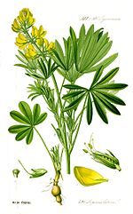 Lupinus luteus (yellow lupine) Lupinus luteus (yellow lupine) | ||||||
| Method |  X-RAY DIFFRACTION / SYNCHROTRON / MOLECULAR REPLACEMENT / Resolution: 1.35 Å X-RAY DIFFRACTION / SYNCHROTRON / MOLECULAR REPLACEMENT / Resolution: 1.35 Å | ||||||
 Authors Authors | Fernandes, H.C. / Pasternak, O. / Bujacz, G. / Bujacz, A. / Sikorski, M.M. / Jaskolski, M. | ||||||
 Citation Citation | Journal: J.Mol.Biol. / Year: 2008 Title: Lupinus luteus pathogenesis-related protein as a reservoir for cytokinin. Authors: Fernandes, H. / Pasternak, O. / Bujacz, G. / Bujacz, A. / Sikorski, M.M. / Jaskolski, M. #1: Journal: Acta Crystallogr.,Sect.D / Year: 2005Title: Structure of a yellow lupin pathogenesis-related PR-10 protein belonging to a novel subclass Authors: Pasternak, O. / Biesiadka, J. / Dolot, R. / Handschuh, L. / Bujacz, G. / Sikorski, M.M. / Jaskolski, M. #2: Journal: J.Mol.Biol. / Year: 2002Title: Crystal structures of two homologous pathogenesis-related proteins from yellow lupine Authors: Biesiadka, J. / Bujacz, G. / Sikorski, M.M. / Jaskolski, M. #3: Journal: Plant cell / Year: 2006Title: Crystal Structure of Vigna radiata Cytokinin-Specific Binding Protein in Complex with Zeatin Authors: Pasternak, O. / Bujacz, G.D. / Fujimoto, Y. / Hashimoto, Y. / Jelen, F. / Otlewski, J. / Sikorski, M.M. / Jaskolski, M. #4: Journal: Nat.Struct.Biol. / Year: 1996Title: X-ray and NMR structure of Bet v 1, the origin of birch pollen allergy Authors: Gajhede, M. / Osmark, P. / Poulsen, F.M. / Ipsen, H. / Larsen, J.N. / Joost van Neerven, R.J. / Schou, C. / Lowenstein, H. / Spangfort, M.D. #5: Journal: J.Mol.Biol. / Year: 2003Title: Crystal structure of a hypoallergenic isoform of the major birch pollen allergen Bet v 1 and its likely biological function as a plant steroid carrier. Authors: Markovic-Housley, Z. / Degano, M. / Lamba, D. / von Roepenack-Lahaye, E. / Clemens, S. / Susani, M. / Ferreira, F. / Scheiner, O. / Breiteneder, H. | ||||||
| History |
|
- Structure visualization
| Structure viewer | Molecule: MolmilJmol/JSmol |
|---|
- Downloads & links
-Download
| PDBx/mmCIF format | 2qim.cif.gz | 89.4 KB | Display | PDBx/mmCIF format |
|---|---|---|---|---|
| PDB format | pdb2qim.ent.gz | 69 KB | Display | PDB format |
| PDBx/mmJSON format | 2qim.json.gz | Tree view | PDBx/mmJSON format | |
| Others |  Other downloads Other downloads |
-Validation report
| Arichive directory | https://data.pdbj.org/pub/pdb/validation_reports/qi/2qimftp://data.pdbj.org/pub/pdb/validation_reports/qi/2qim | HTTPS FTP |
|---|
-Related structure data
| Related structure data | |
|---|---|
| Similar structure data |


-Links
 PDBj
PDBj- Assembly
| Deposited unit | 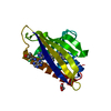
| ||||||||
|---|---|---|---|---|---|---|---|---|---|
| 1 |
| ||||||||
| Unit cell |
|
-Components
| #1: Protein | Mass: 16906.000 Da / Num. of mol.: 1 Source method: isolated from a genetically manipulated source Source: (gene. exp.) Lupinus luteus (yellow lupine) / Gene: pr10.2b, Ypr10.2b / Plasmid: pET3a / Production host:  | ||||
|---|---|---|---|---|---|
| #2: Chemical | ChemComp-CA /   Mass: 40.078 Da / Num. of mol.: 1 / Source method: obtained synthetically / Formula: Ca Mass: 40.078 Da / Num. of mol.: 1 / Source method: obtained synthetically / Formula: Ca | ||||
| #3: Chemical | ChemComp-ZEA / (   Mass: 219.243 Da / Num. of mol.: 4 / Source method: obtained synthetically / Formula: C10H13N5O Mass: 219.243 Da / Num. of mol.: 4 / Source method: obtained synthetically / Formula: C10H13N5O#4: Chemical | ChemComp-GOL / |   Mass: 92.094 Da / Num. of mol.: 1 / Source method: obtained synthetically / Formula: C3H8O3 Mass: 92.094 Da / Num. of mol.: 1 / Source method: obtained synthetically / Formula: C3H8O3#5: Water | ChemComp-HOH / |  Mass: 18.015 Da / Num. of mol.: 233 / Source method: isolated from a natural source / Formula: H2O Mass: 18.015 Da / Num. of mol.: 233 / Source method: isolated from a natural source / Formula: H2O |
-Experimental details
-Experiment
| Experiment | Method: X-RAY DIFFRACTION / Number of used crystals: 1 |
|---|
- Sample preparation
| Crystal | Density Matthews: 3.13 Å3/Da / Density % sol: 60.65 % |
|---|---|
| Crystal grow | Temperature: 292 K / Method: vapor diffusion, hanging drop / pH: 6.5 Details: 1.2 M sodium citrate, 0.1 M MES pH 6.5, VAPOR DIFFUSION, HANGING DROP, temperature 292K |
-Data collection
| Diffraction | Mean temperature: 100 K |
|---|---|
| Diffraction source | Source: SYNCHROTRON / Site: EMBL/DESY, HAMBURG  / Beamline: X13 / Wavelength: 0.803 Å / Beamline: X13 / Wavelength: 0.803 Å |
| Detector | Type: MARRESEARCH / Detector: CCD / Date: Jan 23, 2005 / Details: mirrors |
| Radiation | Monochromator: Si[111], horizontally focussing / Protocol: SINGLE WAVELENGTH / Monochromatic (M) / Laue (L): M / Scattering type: x-ray |
| Radiation wavelength | Wavelength: 0.803 Å / Relative weight: 1 |
| Reflection | Resolution: 1.35→15 Å / Num. all: 45685 / Num. obs: 45644 / % possible obs: 100 % / Observed criterion σ(F): 0 / Observed criterion σ(I): -1 / Redundancy: 9.7 % / Rmerge(I) obs: 0.042 / Net I/σ(I): 56.7 |
| Reflection shell | Resolution: 1.35→1.4 Å / Redundancy: 10.1 % / Rmerge(I) obs: 0.67 / Mean I/σ(I) obs: 2.5 / % possible all: 100 |
- Processing
| Software |
| ||||||||||||||||||||||||||||||||||||||||||||||||||||||||||||||||||||||||||||||||||||||||||||||||||||||||||||||||||||||||||||||||||||||||||||||||||||||||||||||||||||||||||
|---|---|---|---|---|---|---|---|---|---|---|---|---|---|---|---|---|---|---|---|---|---|---|---|---|---|---|---|---|---|---|---|---|---|---|---|---|---|---|---|---|---|---|---|---|---|---|---|---|---|---|---|---|---|---|---|---|---|---|---|---|---|---|---|---|---|---|---|---|---|---|---|---|---|---|---|---|---|---|---|---|---|---|---|---|---|---|---|---|---|---|---|---|---|---|---|---|---|---|---|---|---|---|---|---|---|---|---|---|---|---|---|---|---|---|---|---|---|---|---|---|---|---|---|---|---|---|---|---|---|---|---|---|---|---|---|---|---|---|---|---|---|---|---|---|---|---|---|---|---|---|---|---|---|---|---|---|---|---|---|---|---|---|---|---|---|---|---|---|---|---|---|
| Refinement | Method to determine structure: MOLECULAR REPLACEMENT Starting model: LlPR-10.2F Resolution: 1.35→15 Å / Cor.coef. Fo:Fc: 0.975 / Cor.coef. Fo:Fc free: 0.961 / SU B: 1.642 / SU ML: 0.03 Isotropic thermal model: Anisotropic displacement parameters for non-H atoms Cross valid method: THROUGHOUT / ESU R: 0.044 / ESU R Free: 0.049 / Stereochemistry target values: MAXIMUM LIKELIHOOD / Details: HYDROGENS HAVE BEEN ADDED IN THE RIDING POSITIONS
| ||||||||||||||||||||||||||||||||||||||||||||||||||||||||||||||||||||||||||||||||||||||||||||||||||||||||||||||||||||||||||||||||||||||||||||||||||||||||||||||||||||||||||
| Solvent computation | Ion probe radii: 0.8 Å / Shrinkage radii: 0.8 Å / VDW probe radii: 1.4 Å / Solvent model: BABINET MODEL WITH MASK | ||||||||||||||||||||||||||||||||||||||||||||||||||||||||||||||||||||||||||||||||||||||||||||||||||||||||||||||||||||||||||||||||||||||||||||||||||||||||||||||||||||||||||
| Displacement parameters | Biso mean: 19.218 Å2
| ||||||||||||||||||||||||||||||||||||||||||||||||||||||||||||||||||||||||||||||||||||||||||||||||||||||||||||||||||||||||||||||||||||||||||||||||||||||||||||||||||||||||||
| Refinement step | Cycle: LAST / Resolution: 1.35→15 Å
| ||||||||||||||||||||||||||||||||||||||||||||||||||||||||||||||||||||||||||||||||||||||||||||||||||||||||||||||||||||||||||||||||||||||||||||||||||||||||||||||||||||||||||
| Refine LS restraints |
| ||||||||||||||||||||||||||||||||||||||||||||||||||||||||||||||||||||||||||||||||||||||||||||||||||||||||||||||||||||||||||||||||||||||||||||||||||||||||||||||||||||||||||
| LS refinement shell | Resolution: 1.35→1.385 Å / Total num. of bins used: 20
| ||||||||||||||||||||||||||||||||||||||||||||||||||||||||||||||||||||||||||||||||||||||||||||||||||||||||||||||||||||||||||||||||||||||||||||||||||||||||||||||||||||||||||
| Refinement TLS params. | Method: refined / Origin x: 63.4287 Å / Origin y: 48.9393 Å / Origin z: 0.2718 Å
|
