- PDB-2ope: Crystal structure of the Neisseria meningitidis minor Type IV pil... -
+
Open data
ID or keywords:
Loading...
-
Basic information
Entry
Database: PDB / ID: 2ope
Title
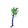
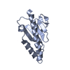
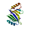
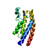
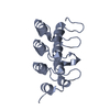
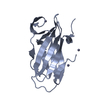
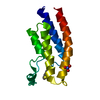
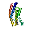
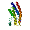
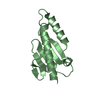
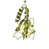
Crystal structure of the Neisseria meningitidis minor Type IV pilin, PilX, in space group P43
Components
PilX
Keywords
CELL ADHESION / Neisseria meningitidis / type IV pilin / PilX / minor pilin / bacterial pathogenesis / adhesion / aggregation / filament
Function / homology
Function and homology information
protein secretion by the type II secretion system / type II protein secretion system complex / membrane Similarity search - Function
Fructose-1,6-Bisphosphatase; Chain A, domain 1 - #20 / Minor type IV pilin, PilX-like / Minor type IV pilin, PilX-like superfamily / Minor type IV pilin, PilX / Bacterial general secretion pathway protein G-type pilin / Prokaryotic N-terminal methylation site. / Prokaryotic N-terminal methylation motif / Prokaryotic N-terminal methylation site / Pilin-like / Fructose-1,6-Bisphosphatase; Chain A, domain 1 ...Fructose-1,6-Bisphosphatase; Chain A, domain 1 - #20 / Minor type IV pilin, PilX-like / Minor type IV pilin, PilX-like superfamily / Minor type IV pilin, PilX / Bacterial general secretion pathway protein G-type pilin / Prokaryotic N-terminal methylation site. / Prokaryotic N-terminal methylation motif / Prokaryotic N-terminal methylation site / Pilin-like / Fructose-1,6-Bisphosphatase; Chain A, domain 1 / 2-Layer Sandwich / Alpha Beta Similarity search - Domain/homology
Type: BRUKER PROTEUM / Detector: CCD / Date: May 18, 2005 / Details: Montel 200
Radiation
Monochromator: graded multilayer mirrors / Protocol: SINGLE WAVELENGTH / Monochromatic (M) / Laue (L): M / Scattering type: x-ray
Radiation wavelength
Wavelength: 1.5418 Å / Relative weight: 1
Reflection
Resolution: 2.4→25 Å / Num. obs: 20280 / % possible obs: 98.6 % / Redundancy: 6.4 % / Rmerge(I) obs: 0.072 / Χ2: 0.964 / Net I/σ(I): 9.7
Reflection shell
Resolution (Å)
Redundancy (%)
Rmerge(I) obs
Num. unique all
Χ2
% possible all
2.4-2.49
3.1
0.243
1794
0.71
87.2
2.49-2.59
3.8
0.218
2008
0.73
98.4
2.59-2.7
5.4
0.18
2044
0.793
100
2.7-2.84
7.2
0.165
2039
0.82
100
2.84-3.02
7.4
0.14
2060
0.934
100
3.02-3.26
7.4
0.107
2030
1.036
100
3.26-3.58
7.4
0.072
2062
1.05
100
3.58-4.1
7.4
0.057
2058
1.024
100
4.1-5.16
7.4
0.046
2079
0.976
100
5.16-25
7.2
0.055
2106
1.19
100
-
Processing
Software
Name
Version
Classification
NB
DENZO
datareduction
SCALEPACK
datascaling
REFMAC
5.2.0019
refinement
PDB_EXTRACT
2
dataextraction
PROTEUM PLUS
PLUS
datacollection
HKL-2000
datareduction
SOLVE
forsadphasing
phasing
RESOLVE
forsadphasing
phasing
Refinement
Method to determine structure: Combination of SAD, Mol. Replacement Resolution: 2.4→25 Å / Cor.coef. Fo:Fc: 0.947 / Cor.coef. Fo:Fc free: 0.915 / SU B: 16.586 / SU ML: 0.205 / Cross valid method: THROUGHOUT / ESU R: 0.504 / ESU R Free: 0.305 / Stereochemistry target values: MAXIMUM LIKELIHOOD Details: The P43 structure was initially solved by SAD phasing using SeMet-substituted PilX. This initial model was used for molecular replacement to solve the companion C2221 structure (2OPD) for ...Details: The P43 structure was initially solved by SAD phasing using SeMet-substituted PilX. This initial model was used for molecular replacement to solve the companion C2221 structure (2OPD) for which the data are substantially better. After that structure was refined, it was used as the molecular replacement model for structure solution of the P43 structure using native data collected on a rotating anode.
Rfactor
Num. reflection
% reflection
Selection details
Rfree
0.27893
1033
5.1 %
RANDOM
Rwork
0.20794
-
-
-
obs
0.21156
19111
98.61 %
-
Solvent computation
Ion probe radii: 0.8 Å / Shrinkage radii: 0.8 Å / VDW probe radii: 1.4 Å / Solvent model: BABINET MODEL WITH MASK
Displacement parameters
Biso mean: 47.078 Å2
Baniso -1
Baniso -2
Baniso -3
1-
0.05 Å2
0 Å2
0 Å2
2-
-
0.05 Å2
0 Å2
3-
-
-
-0.1 Å2
Refine analyze
Luzzati coordinate error free: 0.303 Å
Refinement step
Cycle: LAST / Resolution: 2.4→25 Å
Protein
Nucleic acid
Ligand
Solvent
Total
Num. atoms
3622
0
0
181
3803
Refine LS restraints
Refine-ID
Type
Dev ideal
Dev ideal target
Number
X-RAY DIFFRACTION
r_bond_refined_d
0.018
0.022
3693
X-RAY DIFFRACTION
r_bond_other_d
X-RAY DIFFRACTION
r_angle_refined_deg
1.486
1.954
4976
X-RAY DIFFRACTION
r_angle_other_deg
X-RAY DIFFRACTION
r_dihedral_angle_1_deg
6.603
5
457
X-RAY DIFFRACTION
r_dihedral_angle_2_deg
36.091
24.534
161
X-RAY DIFFRACTION
r_dihedral_angle_3_deg
15.934
15
651
X-RAY DIFFRACTION
r_dihedral_angle_4_deg
16.753
15
16
X-RAY DIFFRACTION
r_chiral_restr
0.111
0.2
543
X-RAY DIFFRACTION
r_gen_planes_refined
0.006
0.02
2748
X-RAY DIFFRACTION
r_gen_planes_other
X-RAY DIFFRACTION
r_nbd_refined
0.233
0.3
1839
X-RAY DIFFRACTION
r_nbd_other
X-RAY DIFFRACTION
r_nbtor_refined
0.319
0.5
2540
X-RAY DIFFRACTION
r_nbtor_other
X-RAY DIFFRACTION
r_xyhbond_nbd_refined
0.228
0.5
345
X-RAY DIFFRACTION
r_xyhbond_nbd_other
X-RAY DIFFRACTION
r_metal_ion_refined
X-RAY DIFFRACTION
r_metal_ion_other
X-RAY DIFFRACTION
r_symmetry_vdw_refined
0.265
0.3
76
X-RAY DIFFRACTION
r_symmetry_vdw_other
X-RAY DIFFRACTION
r_symmetry_hbond_refined
0.189
0.5
12
X-RAY DIFFRACTION
r_symmetry_hbond_other
X-RAY DIFFRACTION
r_symmetry_metal_ion_refined
X-RAY DIFFRACTION
r_symmetry_metal_ion_other
X-RAY DIFFRACTION
r_mcbond_it
1.027
2
2349
X-RAY DIFFRACTION
r_mcbond_other
X-RAY DIFFRACTION
r_mcangle_it
1.598
3
3679
X-RAY DIFFRACTION
r_scbond_it
1.159
2
1538
X-RAY DIFFRACTION
r_scangle_it
1.634
3
1297
X-RAY DIFFRACTION
r_rigid_bond_restr
X-RAY DIFFRACTION
r_sphericity_free
X-RAY DIFFRACTION
r_sphericity_bonded
LS refinement shell
Resolution: 2.4→2.462 Å / Total num. of bins used: 20
Rfactor
Num. reflection
% reflection
Rfree
0.29
68
-
Rwork
0.217
1201
-
obs
-
1269
86.09 %
+
About Yorodumi
-
News
-
Feb 9, 2022. New format data for meta-information of EMDB entries
New format data for meta-information of EMDB entries
Version 3 of the EMDB header file is now the official format.
The previous official version 1.9 will be removed from the archive.
In the structure databanks used in Yorodumi, some data are registered as the other names, "COVID-19 virus" and "2019-nCoV". Here are the details of the virus and the list of structure data.
Jan 31, 2019. EMDB accession codes are about to change! (news from PDBe EMDB page)
EMDB accession codes are about to change! (news from PDBe EMDB page)
The allocation of 4 digits for EMDB accession codes will soon come to an end. Whilst these codes will remain in use, new EMDB accession codes will include an additional digit and will expand incrementally as the available range of codes is exhausted. The current 4-digit format prefixed with “EMD-” (i.e. EMD-XXXX) will advance to a 5-digit format (i.e. EMD-XXXXX), and so on. It is currently estimated that the 4-digit codes will be depleted around Spring 2019, at which point the 5-digit format will come into force.
The EM Navigator/Yorodumi systems omit the EMD- prefix.
Related info.:Q: What is EMD? / ID/Accession-code notation in Yorodumi/EM Navigator
Yorodumi is a browser for structure data from EMDB, PDB, SASBDB, etc.
This page is also the successor to EM Navigator detail page, and also detail information page/front-end page for Omokage search.
The word "yorodu" (or yorozu) is an old Japanese word meaning "ten thousand". "mi" (miru) is to see.
Related info.:EMDB / PDB / SASBDB / Comparison of 3 databanks / Yorodumi Search / Aug 31, 2016. New EM Navigator & Yorodumi / Yorodumi Papers / Jmol/JSmol / Function and homology information / Changes in new EM Navigator and Yorodumi
 Movie
Movie Controller

 Yorodumi
 Open data
 Basic information
 Components
 Keywords
 CELL ADHESION /
  Function and homology information
 Authors
 Citation
 Structure visualization
 Downloads & links
 Other downloads


 PDBj
PDBj
 Assembly
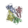


 Mass: 18.015 Da / Num. of mol.: 181 / Source method: isolated from a natural source / Formula: H2O
 Sample preparation
 Processing