 Movie
Movie Controller

[English] 日本語
 Yorodumi
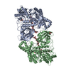
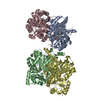
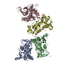
- PDB-2oat: ORNITHINE AMINOTRANSFERASE COMPLEXED WITH 5-FLUOROMETHYLORNITHINE -
+ Open data

- Basic information
| Entry | Database: PDB / ID: 2oat | ||||||
|---|---|---|---|---|---|---|---|
| Title | ORNITHINE AMINOTRANSFERASE COMPLEXED WITH 5-FLUOROMETHYLORNITHINE | ||||||
 Components Components | ORNITHINE AMINOTRANSFERASE | ||||||
 Keywords Keywords | AMINOTRANSFERASE / 5-FLUOROMETHYLORNITHINE / PLP-DEPENDENT ENZYME / PYRIDOXAL PHOSPHATE | ||||||
| Function / homology |  Function and homology information Function and homology informationarginine catabolic process to proline via ornithine / ornithine aminotransferase activity / ornithine aminotransferase / arginine catabolic process to glutamate / L-proline biosynthetic process / Glutamate and glutamine metabolism / visual perception / pyridoxal phosphate binding / mitochondrial matrix / mitochondrion ...arginine catabolic process to proline via ornithine / ornithine aminotransferase activity / ornithine aminotransferase / arginine catabolic process to glutamate / L-proline biosynthetic process / Glutamate and glutamine metabolism / visual perception / pyridoxal phosphate binding / mitochondrial matrix / mitochondrion / nucleoplasm / identical protein binding / cytoplasm Similarity search - Function | ||||||
| Biological species |  Homo sapiens (human) Homo sapiens (human) | ||||||
| Method |  X-RAY DIFFRACTION / SYNCHROTRON / DIFFERENCE FOURIER TECHNIQUES / Resolution: 1.95 Å X-RAY DIFFRACTION / SYNCHROTRON / DIFFERENCE FOURIER TECHNIQUES / Resolution: 1.95 Å | ||||||
 Authors Authors | Storici, P. / Schirmer, T. | ||||||
 Citation Citation | Journal: J.Mol.Biol. / Year: 1999 Title: Crystal structure of human ornithine aminotransferase complexed with the highly specific and potent inhibitor 5-fluoromethylornithine. Authors: Storici, P. / Capitani, G. / Muller, R. / Schirmer, T. / Jansonius, J.N. #1: Journal: J.Mol.Biol. / Year: 1998Title: Crystal Structure of Human Recombinant Ornithine Aminotransferase Authors: Shen, B.W. / Hennig, M. / Hohenester, E. / Jansonius, J.N. / Schirmer, T. #2: Journal: J.Mol.Biol. / Year: 1994Title: Crystallization and Preliminary X-Ray Diffraction Studies of Recombinant Human Ornithine Aminotransferase Authors: Shen, B.W. / Ramesh, V. / Mueller, R. / Hohenester, E. / Hennig, M. / Jansonius, J.N. #3: Journal: Biochem.J. / Year: 1990Title: Dl-Canaline and 5-Fluoromethylornithine. Comparison of Two Inactivators of Ornithine Aminotransferase Authors: Bolkenius, F.N. / Knodgen, B. / Seiler, N. | ||||||
| History |
|
- Structure visualization
| Structure viewer | Molecule: MolmilJmol/JSmol |
|---|
- Downloads & links
-Download
| PDBx/mmCIF format | 2oat.cif.gz | 256.8 KB | Display | PDBx/mmCIF format |
|---|---|---|---|---|
| PDB format | pdb2oat.ent.gz | 206.9 KB | Display | PDB format |
| PDBx/mmJSON format | 2oat.json.gz | Tree view | PDBx/mmJSON format | |
| Others |  Other downloads Other downloads |
-Validation report
| Summary document | 2oat_validation.pdf.gz | 545.8 KB | Display | wwPDB validaton report |
|---|---|---|---|---|
| Full document | 2oat_full_validation.pdf.gz | 571 KB | Display | |
| Data in XML | 2oat_validation.xml.gz | 27.6 KB | Display | |
| Data in CIF | 2oat_validation.cif.gz | 45 KB | Display | |
| Arichive directory | https://data.pdbj.org/pub/pdb/validation_reports/oa/2oatftp://data.pdbj.org/pub/pdb/validation_reports/oa/2oat | HTTPS FTP |
-Related structure data
| Related structure data | 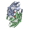 1oatS S: Starting model for refinement |
|---|---|
| Similar structure data |


-Links
 PDBj
PDBj- Assembly
| Deposited unit | 
| ||||||||||||
|---|---|---|---|---|---|---|---|---|---|---|---|---|---|
| 1 | 
| ||||||||||||
| 2 | 
| ||||||||||||
| 3 | 
| ||||||||||||
| Unit cell |
| ||||||||||||
| Noncrystallographic symmetry (NCS) | NCS oper:
| ||||||||||||
| Details | THERE ARE ONE AND A HALF DIMERS IN THE ASYMMETRIC UNIT. CHAINS A AND B REFER TO THE TWO SUBUNITS OF THE FIRST DIMER. CHAIN C REFERS TO ONE SUBUNIT OF THE SECOND DIMER; THE OTHER SUBUNIT IS RELATED BY CRYSTAL SYMMETRY. |
-Components
| #1: Protein | Mass: 48593.668 Da / Num. of mol.: 3 Source method: isolated from a genetically manipulated source Source: (gene. exp.) Homo sapiens (human) / Cellular location: INTRAMITOCHONDRIA / Gene: OAT / Organ: LIVER / Organelle: MITOCHONDRIA / Plasmid: PMAL-C2 / Production host:  #2: Chemical |   Mass: 374.283 Da / Num. of mol.: 3 / Source method: obtained synthetically / Formula: C14H19N2O8P Mass: 374.283 Da / Num. of mol.: 3 / Source method: obtained synthetically / Formula: C14H19N2O8P#3: Water | ChemComp-HOH / |  Mass: 18.015 Da / Num. of mol.: 744 / Source method: isolated from a natural source / Formula: H2O Mass: 18.015 Da / Num. of mol.: 744 / Source method: isolated from a natural source / Formula: H2O |
|---|
-Experimental details
-Experiment
| Experiment | Method: X-RAY DIFFRACTION / Number of used crystals: 1 |
|---|
- Sample preparation
| Crystal | Density Matthews: 2.65 Å3/Da / Density % sol: 50.9 % | ||||||||||||||||||||||||||||||||||||
|---|---|---|---|---|---|---|---|---|---|---|---|---|---|---|---|---|---|---|---|---|---|---|---|---|---|---|---|---|---|---|---|---|---|---|---|---|---|
| Crystal grow | pH: 7.9 Details: (2S,5S)5FMORN-OAT WAS CO-CRYSTALLIZED FROM 6-10% PEG 6000, 1MM DTT, 120-160 MM NACL, 10-20% GLYCEROL, 50 MM TRICIN, PH 7.9. | ||||||||||||||||||||||||||||||||||||
| Crystal grow | *PLUS Method: vapor diffusion, hanging drop | ||||||||||||||||||||||||||||||||||||
| Components of the solutions | *PLUS
|
-Data collection
| Diffraction | Mean temperature: 90 K |
|---|---|
| Diffraction source | Source: SYNCHROTRON / Site: MPG/DESY, HAMBURG  / Beamline: BW6 / Wavelength: 1.1 / Beamline: BW6 / Wavelength: 1.1 |
| Detector | Type: MARRESEARCH / Detector: IMAGE PLATE / Date: Nov 1, 1996 |
| Radiation | Monochromator: GRAPHITE(002) / Monochromatic (M) / Laue (L): M / Scattering type: x-ray |
| Radiation wavelength | Wavelength: 1.1 Å / Relative weight: 1 |
| Reflection | Resolution: 1.95→19.9 Å / Num. obs: 101836 / % possible obs: 97 % / Observed criterion σ(I): 0 / Redundancy: 3.2 % / Rsym value: 0.076 / Net I/σ(I): 12.5 |
| Reflection shell | Resolution: 1.95→1.98 Å / Mean I/σ(I) obs: 3.4 / Rsym value: 0.35 / % possible all: 94 |
| Reflection | *PLUS Rmerge(I) obs: 0.076 |
| Reflection shell | *PLUS % possible obs: 94 % / Rmerge(I) obs: 0.297 |
- Processing
| Software |
| |||||||||||||||||||||||||||||||||||||||||||||||||||||||||||||||
|---|---|---|---|---|---|---|---|---|---|---|---|---|---|---|---|---|---|---|---|---|---|---|---|---|---|---|---|---|---|---|---|---|---|---|---|---|---|---|---|---|---|---|---|---|---|---|---|---|---|---|---|---|---|---|---|---|---|---|---|---|---|---|---|---|
| Refinement | Method to determine structure: DIFFERENCE FOURIER TECHNIQUES Starting model: 1OAT Resolution: 1.95→19.6 Å / Cross valid method: THROUGHOUT / σ(F): 0
| |||||||||||||||||||||||||||||||||||||||||||||||||||||||||||||||
| Displacement parameters | Biso mean: 25.2 Å2 | |||||||||||||||||||||||||||||||||||||||||||||||||||||||||||||||
| Refinement step | Cycle: LAST / Resolution: 1.95→19.6 Å
| |||||||||||||||||||||||||||||||||||||||||||||||||||||||||||||||
| Refine LS restraints |
| |||||||||||||||||||||||||||||||||||||||||||||||||||||||||||||||
| Software | *PLUS Name: REFMAC / Classification: refinement | |||||||||||||||||||||||||||||||||||||||||||||||||||||||||||||||
| Refinement | *PLUS Lowest resolution: 19.5 Å | |||||||||||||||||||||||||||||||||||||||||||||||||||||||||||||||
| Solvent computation | *PLUS | |||||||||||||||||||||||||||||||||||||||||||||||||||||||||||||||
| Displacement parameters | *PLUS | |||||||||||||||||||||||||||||||||||||||||||||||||||||||||||||||
| Refine LS restraints | *PLUS
|
