[English] 日本語
Yorodumi
- PDB-3v21: Crystal structure of Type IIF restriction endonuclease Bse634I wi... -

+
Open data


ID or keywords:

Loading...

-
Basic information

Entry
Database: PDB / ID: 3v21
TitleCrystal structure of Type IIF restriction endonuclease Bse634I with cognate DNA
Components
  • DNA (5'-D(*TP*TP*CP*GP*AP*CP*CP*GP*GP*TP*CP*GP*A)-3')
  • Endonuclease Bse634IR
KeywordsDNA BINDING PROTEIN / HYDROLASE/DNA / RESTRICTION ENDONUCLEASE / protein-DNA complex / HYDROLASE / HYDROLASE-DNA complex
Function / homology
Function and homology information


endonuclease activity / metal ion binding
Similarity search - Function
Restriction Endonuclease - #10 / Restriction endonuclease, type II, Cfr10I/Bse634I / Cfr10I/Bse634I restriction endonuclease / Restriction Endonuclease / Restriction endonuclease type II-like / 3-Layer(aba) Sandwich / Alpha Beta
Similarity search - Domain/homology
DNA / DNA (> 10) / Endonuclease Bse634IR
Similarity search - Component
Biological speciesGeobacillus stearothermophilus (bacteria)
MethodX-RAY DIFFRACTION / SYNCHROTRON / molecular replacement / Resolution: 2.7 Å
AuthorsManakova, E.N. / Grazulis, S. / Golovenko, D. / Tamulaitiene, G.
CitationJournal: Nucleic Acids Res. / Year: 2012
Title: Structural mechanisms of the degenerate sequence recognition by Bse634I restriction endonuclease.
Authors: Manakova, E. / Grazulis, S. / Zaremba, M. / Tamulaitiene, G. / Golovenko, D. / Siksnys, V.
History
DepositionDec 11, 2011Deposition site: RCSB / Processing site: RCSB
Revision 1.0Apr 25, 2012Provider: repository / Type: Initial release
Revision 1.1Nov 14, 2012Group: Database references
Revision 1.2Sep 13, 2023Group: Data collection / Database references / Refinement description
Category: chem_comp_atom / chem_comp_bond ...chem_comp_atom / chem_comp_bond / database_2 / diffrn_source / pdbx_initial_refinement_model / struct_ref_seq_dif
Item: _database_2.pdbx_DOI / _database_2.pdbx_database_accession ..._database_2.pdbx_DOI / _database_2.pdbx_database_accession / _diffrn_source.pdbx_synchrotron_site / _struct_ref_seq_dif.details

-
Structure visualization

Structure viewerMolecule:
MolmilJmol/JSmol

Downloads & links

-
Assembly

Deposited unit
A: Endonuclease Bse634IR
B: Endonuclease Bse634IR
C: Endonuclease Bse634IR
D: Endonuclease Bse634IR
E: Endonuclease Bse634IR
F: Endonuclease Bse634IR
G: Endonuclease Bse634IR
H: Endonuclease Bse634IR
I: DNA (5'-D(*TP*TP*CP*GP*AP*CP*CP*GP*GP*TP*CP*GP*A)-3')
J: DNA (5'-D(*TP*TP*CP*GP*AP*CP*CP*GP*GP*TP*CP*GP*A)-3')
K: DNA (5'-D(*TP*TP*CP*GP*AP*CP*CP*GP*GP*TP*CP*GP*A)-3')
L: DNA (5'-D(*TP*TP*CP*GP*AP*CP*CP*GP*GP*TP*CP*GP*A)-3')
M: DNA (5'-D(*TP*TP*CP*GP*AP*CP*CP*GP*GP*TP*CP*GP*A)-3')
N: DNA (5'-D(*TP*TP*CP*GP*AP*CP*CP*GP*GP*TP*CP*GP*A)-3')
O: DNA (5'-D(*TP*TP*CP*GP*AP*CP*CP*GP*GP*TP*CP*GP*A)-3')
P: DNA (5'-D(*TP*TP*CP*GP*AP*CP*CP*GP*GP*TP*CP*GP*A)-3')
R: DNA (5'-D(*TP*TP*CP*GP*AP*CP*CP*GP*GP*TP*CP*GP*A)-3')
S: DNA (5'-D(*TP*TP*CP*GP*AP*CP*CP*GP*GP*TP*CP*GP*A)-3')
V: DNA (5'-D(*TP*TP*CP*GP*AP*CP*CP*GP*GP*TP*CP*GP*A)-3')
X: DNA (5'-D(*TP*TP*CP*GP*AP*CP*CP*GP*GP*TP*CP*GP*A)-3')
Y: DNA (5'-D(*TP*TP*CP*GP*AP*CP*CP*GP*GP*TP*CP*GP*A)-3')
Z: DNA (5'-D(*TP*TP*CP*GP*AP*CP*CP*GP*GP*TP*CP*GP*A)-3')


Theoretical massNumber of molelcules
Total (without water)325,64822
Polymers325,64822
Non-polymers00
Water6,467359
1
A: Endonuclease Bse634IR
B: Endonuclease Bse634IR
E: Endonuclease Bse634IR
F: Endonuclease Bse634IR
I: DNA (5'-D(*TP*TP*CP*GP*AP*CP*CP*GP*GP*TP*CP*GP*A)-3')
J: DNA (5'-D(*TP*TP*CP*GP*AP*CP*CP*GP*GP*TP*CP*GP*A)-3')
M: DNA (5'-D(*TP*TP*CP*GP*AP*CP*CP*GP*GP*TP*CP*GP*A)-3')
N: DNA (5'-D(*TP*TP*CP*GP*AP*CP*CP*GP*GP*TP*CP*GP*A)-3')


Theoretical massNumber of molelcules
Total (without water)150,9218
Polymers150,9218
Non-polymers00
Water1448
TypeNameSymmetry operationNumber
identity operation1_555x,y,z1
Buried area23220 Å2
ΔGint-128 kcal/mol
Surface area48600 Å2
MethodPISA
2
C: Endonuclease Bse634IR
D: Endonuclease Bse634IR
G: Endonuclease Bse634IR
H: Endonuclease Bse634IR
K: DNA (5'-D(*TP*TP*CP*GP*AP*CP*CP*GP*GP*TP*CP*GP*A)-3')
L: DNA (5'-D(*TP*TP*CP*GP*AP*CP*CP*GP*GP*TP*CP*GP*A)-3')
O: DNA (5'-D(*TP*TP*CP*GP*AP*CP*CP*GP*GP*TP*CP*GP*A)-3')
P: DNA (5'-D(*TP*TP*CP*GP*AP*CP*CP*GP*GP*TP*CP*GP*A)-3')


Theoretical massNumber of molelcules
Total (without water)150,9218
Polymers150,9218
Non-polymers00
Water1448
TypeNameSymmetry operationNumber
identity operation1_555x,y,z1
Buried area23200 Å2
ΔGint-125 kcal/mol
Surface area48420 Å2
MethodPISA
3
V: DNA (5'-D(*TP*TP*CP*GP*AP*CP*CP*GP*GP*TP*CP*GP*A)-3')
Z: DNA (5'-D(*TP*TP*CP*GP*AP*CP*CP*GP*GP*TP*CP*GP*A)-3')


Theoretical massNumber of molelcules
Total (without water)7,9352
Polymers7,9352
Non-polymers00
Water362
TypeNameSymmetry operationNumber
identity operation1_555x,y,z1
Buried area1090 Å2
ΔGint-8 kcal/mol
Surface area4670 Å2
MethodPISA
4
X: DNA (5'-D(*TP*TP*CP*GP*AP*CP*CP*GP*GP*TP*CP*GP*A)-3')
Y: DNA (5'-D(*TP*TP*CP*GP*AP*CP*CP*GP*GP*TP*CP*GP*A)-3')


Theoretical massNumber of molelcules
Total (without water)7,9352
Polymers7,9352
Non-polymers00
Water362
TypeNameSymmetry operationNumber
identity operation1_555x,y,z1
Buried area1090 Å2
ΔGint-6 kcal/mol
Surface area4870 Å2
MethodPISA
Unit cell
Length a, b, c (Å)106.880, 115.311, 130.243
Angle α, β, γ (deg.)90.000, 112.500, 90.000
Int Tables number4
Space group name H-MP1211
Details
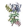
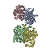
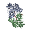
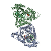
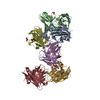
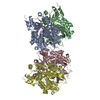
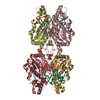
THE BIOLOGICAL ASSEMBLY OF BSE634I IS A TETRAMER FORMED FROM TWO DIMERS STACKED BACK-TO-BACK. EACH PRIMARY DIMER BINDS ONE DNA DUPLEX. THERE ARE TWO TETRAMERS FOUND IN THE ASYMMETRIC UNIT. PROTEIN SUBUNITS AB AND EF FORM THE FIRST TETRAMER, WHICH SPECIFICALLY BINDS TWO DNA DUPLEXES (IJ AND MN). THE SECOND TETRAMER IS COMPOSED FROM TWO DIMERS CD AND GH, EACH DIMER SPECIFICALLY BINDS DNA DUPLEX (KL AND OP). THEREFORE, THERE ARE TWO BIOLOGICAL ASSEMBLIES IN THE ASYM. UNIT. DNA DUPLEXES XY, VZ AND RS ARE NOT PARTS OF THE BIOLOGICAL ASSEMBLY, THEY ARE JUST CRYSTAL PACKING ARTIFACTS.

-
Components

#1: Protein
Endonuclease Bse634IR


Mass: 33762.758 Da / Num. of mol.: 8 / Mutation: R226A
Source method: isolated from a genetically manipulated source
Source: (gene. exp.) Geobacillus stearothermophilus (bacteria)
Strain: 1422 / Gene: bse634IR / Plasmid: pUC18 / Production host: Escherichia coli (E. coli) / Strain (production host): ER2267
References: UniProt: Q8RT53, type II site-specific deoxyribonuclease
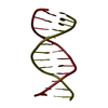
#2: DNA chain
DNA (5'-D(*TP*TP*CP*GP*AP*CP*CP*GP*GP*TP*CP*GP*A)-3')


Mass: 3967.585 Da / Num. of mol.: 14 / Source method: obtained synthetically
#3: Water ChemComp-HOH / water


Mass: 18.015 Da / Num. of mol.: 359 / Source method: isolated from a natural source / Formula: H2O
Sequence detailsSEQUENCE AUTHORS STATE THAT THE RESIDUES AT POSITION 110, 111, AND 130 ARE CORRECTLY IDENTIFIED AND ...SEQUENCE AUTHORS STATE THAT THE RESIDUES AT POSITION 110, 111, AND 130 ARE CORRECTLY IDENTIFIED AND THE BIOCHEMICAL DATA SHOWS THAT THE PROTEIN IS ACTIVE.

-
Experimental details

-
Experiment

ExperimentMethod: X-RAY DIFFRACTION / Number of used crystals: 1

-
Sample preparation

CrystalDensity Matthews: 2.3 Å3/Da / Density % sol: 46.1 %
Crystal growTemperature: 293 K / Method: vapor diffusion / pH: 5.5
Details: 0.1 M Bis-Tris, 0.5% polyvinylpyrrolidone and 16% of PEG400, VAPOR DIFFUSION, temperature 293K, pH 5.5

-
Data collection

DiffractionMean temperature: 100 K
Diffraction sourceSource: SYNCHROTRON / Site: EMBL/DESY, HAMBURG / Beamline: X11 / Wavelength: 0.8148 Å
DetectorType: MAR555 / Detector: CCD / Date: Oct 1, 2007
RadiationMonochromator: Si (111), horizontally focussing / Protocol: SINGLE WAVELENGT / Scattering type: x-ray
Radiation wavelengthWavelength: 0.8148 Å / Relative weight: 1
ReflectionResolution: 2.326→65.094 Å / Num. all: 80299 / Num. obs: 80299 / % possible obs: 100 % / Observed criterion σ(F): 0 / Observed criterion σ(I): 0 / Redundancy: 4.6 % / Biso Wilson estimate: 66.976 Å2 / Rmerge(I) obs: 0.061 / Rsym value: 0.061 / Net I/σ(I): 20.3
Reflection shell

Diffraction-ID: 1

Resolution (Å)Redundancy (%)Rmerge(I) obsMean I/σ(I) obsNum. measured allNum. unique allRsym value% possible all
2.7-2.854.60.3881.953541116960.388100
2.85-3.024.60.2682.750507110260.268100
3.02-3.234.60.1624.447603103950.162100
3.23-3.494.60.0997.24441796860.099100
3.49-3.824.60.06510.64086089230.065100
3.82-4.274.60.04713.93703080830.047100
4.27-4.934.60.03717.33249671070.037100
4.93-6.044.60.03518.72768260700.035100
6.04-8.544.50.0319.52124046850.03100
8.54-65.094.30.02718.31123426280.02799.6

-
Phasing

PhasingMethod: molecular replacement

-
Processing

Software
NameVersionClassificationNB
REFMACrefinement
MOSFLMdata reduction
SCALA3.2.5data scaling
PDB_EXTRACT3.006data extraction
MAR345dtbdata collection
MOLREPphasing
RefinementMethod to determine structure: molecular replacement
Starting model: 1knv
Resolution: 2.7→65.09 Å / Cor.coef. Fo:Fc: 0.918 / Cor.coef. Fo:Fc free: 0.895 / WRfactor Rfree: 0.243 / WRfactor Rwork: 0.209 / Occupancy max: 1 / Occupancy min: 0 / FOM work R set: 0.801 / SU Rfree: 0.405 / Cross valid method: THROUGHOUT / σ(F): 0 / σ(I): 0 / ESU R Free: 0.405 / Stereochemistry target values: MAXIMUM LIKELIHOOD / Details: HYDROGENS HAVE BEEN ADDED IN THE RIDING POSITIONS
RfactorNum. reflection% reflectionSelection details
Rfree0.267 7973 9.9 %RANDOM
Rwork0.23 ---
all0.23372 80279 --
obs0.234 80279 99.98 %-
Solvent computationIon probe radii: 0.8 Å / Shrinkage radii: 0.8 Å / VDW probe radii: 1.4 Å / Solvent model: MASK
Displacement parametersBiso max: 100 Å2 / Biso mean: 54.404 Å2 / Biso min: 13.67 Å2
Baniso -1Baniso -2Baniso -3
1--0.54 Å20 Å2-0.54 Å2
2---0.13 Å20 Å2
3---0.26 Å2
Refinement stepCycle: LAST / Resolution: 2.7→65.09 Å
ProteinNucleic acidLigandSolventTotal
Num. atoms18735 3562 0 359 22656
Refine LS restraints
Refine-IDTypeDev idealDev ideal targetNumber
X-RAY DIFFRACTIONr_bond_refined_d0.0080.02221644
X-RAY DIFFRACTIONr_bond_other_d00.0213800
X-RAY DIFFRACTIONr_angle_refined_deg1.2882.15429951
X-RAY DIFFRACTIONr_angle_other_deg3.82333898
X-RAY DIFFRACTIONr_dihedral_angle_1_deg1.54252175
X-RAY DIFFRACTIONr_dihedral_angle_2_deg39.44224.833836
X-RAY DIFFRACTIONr_dihedral_angle_3_deg15.206153351
X-RAY DIFFRACTIONr_dihedral_angle_4_deg21.6221593
X-RAY DIFFRACTIONr_chiral_restr0.0890.23421
X-RAY DIFFRACTIONr_gen_planes_refined0.0080.0221264
X-RAY DIFFRACTIONr_gen_planes_other0.0050.023458
X-RAY DIFFRACTIONr_nbd_refined0.2440.25567
X-RAY DIFFRACTIONr_nbd_other0.2730.215866
X-RAY DIFFRACTIONr_nbtor_refined0.2030.210567
X-RAY DIFFRACTIONr_nbtor_other0.1070.29860
X-RAY DIFFRACTIONr_xyhbond_nbd_refined0.1560.2691
X-RAY DIFFRACTIONr_xyhbond_nbd_other0.110.25
X-RAY DIFFRACTIONr_symmetry_vdw_refined0.230.247
X-RAY DIFFRACTIONr_symmetry_vdw_other0.3120.2113
X-RAY DIFFRACTIONr_symmetry_hbond_refined0.1150.218
X-RAY DIFFRACTIONr_mcbond_it1.1831.510939
X-RAY DIFFRACTIONr_mcbond_other01.54367
X-RAY DIFFRACTIONr_mcangle_it2.099217787
X-RAY DIFFRACTIONr_scbond_it1.122310705
X-RAY DIFFRACTIONr_scangle_it1.8784.512164
LS refinement shellResolution: 2.7→2.77 Å / Total num. of bins used: 20
RfactorNum. reflection% reflection
Rfree0.417 571 -
Rwork0.358 5322 -
all-5893 -
obs-5322 99.98 %

+
About Yorodumi

-
News

-
Feb 9, 2022. New format data for meta-information of EMDB entries

New format data for meta-information of EMDB entries

  • Version 3 of the EMDB header file is now the official format.
  • The previous official version 1.9 will be removed from the archive.

Related info.:EMDB header

External links:wwPDB to switch to version 3 of the EMDB data model

-
Aug 12, 2020. Covid-19 info

Covid-19 info

URL: https://pdbjlvh1.pdbj.org/emnavi/covid19.php

New page: Covid-19 featured information page in EM Navigator.

Related info.:Covid-19 info / Mar 5, 2020. Novel coronavirus structure data

+
Mar 5, 2020. Novel coronavirus structure data

Novel coronavirus structure data

Related info.:Yorodumi Speices / Aug 12, 2020. Covid-19 info

External links:COVID-19 featured content - PDBj / Molecule of the Month (242):Coronavirus Proteases

+
Jan 31, 2019. EMDB accession codes are about to change! (news from PDBe EMDB page)

EMDB accession codes are about to change! (news from PDBe EMDB page)

  • The allocation of 4 digits for EMDB accession codes will soon come to an end. Whilst these codes will remain in use, new EMDB accession codes will include an additional digit and will expand incrementally as the available range of codes is exhausted. The current 4-digit format prefixed with “EMD-” (i.e. EMD-XXXX) will advance to a 5-digit format (i.e. EMD-XXXXX), and so on. It is currently estimated that the 4-digit codes will be depleted around Spring 2019, at which point the 5-digit format will come into force.
  • The EM Navigator/Yorodumi systems omit the EMD- prefix.

Related info.:Q: What is EMD? / ID/Accession-code notation in Yorodumi/EM Navigator

External links:EMDB Accession Codes are Changing Soon! / Contact to PDBj

+
Jul 12, 2017. Major update of PDB

Major update of PDB

  • wwPDB released updated PDB data conforming to the new PDBx/mmCIF dictionary.
  • This is a major update changing the version number from 4 to 5, and with Remediation, in which all the entries are updated.
  • In this update, many items about electron microscopy experimental information are reorganized (e.g. em_software).
  • Now, EM Navigator and Yorodumi are based on the updated data.

External links:wwPDB Remediation / Enriched Model Files Conforming to OneDep Data Standards Now Available in the PDB FTP Archive

-
Yorodumi

Thousand views of thousand structures

  • Yorodumi is a browser for structure data from EMDB, PDB, SASBDB, etc.
  • This page is also the successor to EM Navigator detail page, and also detail information page/front-end page for Omokage search.
  • The word "yorodu" (or yorozu) is an old Japanese word meaning "ten thousand". "mi" (miru) is to see.

Related info.:EMDB / PDB / SASBDB / Comparison of 3 databanks / Yorodumi Search / Aug 31, 2016. New EM Navigator & Yorodumi / Yorodumi Papers / Jmol/JSmol / Function and homology information / Changes in new EM Navigator and Yorodumi

Read more