 Movie
Movie Controller

[English] 日本語
 Yorodumi
- PDB-2jkp: Structure of a family 97 alpha-glucosidase from Bacteroides theta... -
+ Open data

- Basic information
| Entry | Database: PDB / ID: 2jkp | ||||||
|---|---|---|---|---|---|---|---|
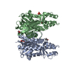
| Title | Structure of a family 97 alpha-glucosidase from Bacteroides thetaiotaomicron in complex with castanospermine | ||||||
 Components Components | ALPHA-GLUCOSIDASE (ALPHA-GLUCOSIDASE SUSB) | ||||||
 Keywords Keywords | HYDROLASE / FAMILY 97 / CASTANOSPERMINE / ALPHA-GLUCOSIDASE / GLYCOSIDE HYDROLASE / BACTEROIDES THETAIOTAOMICRON | ||||||
| Function / homology |  Function and homology information Function and homology informationglucan 1,4-alpha-glucosidase / glucan 1,4-alpha-glucosidase activity / alpha-1,4-glucosidase activity / starch catabolic process / carbohydrate binding / periplasmic space / calcium ion binding / plasma membrane Similarity search - Function | ||||||
| Biological species |  BACTEROIDES THETAIOTAOMICRON (bacteria) BACTEROIDES THETAIOTAOMICRON (bacteria) | ||||||
| Method |  X-RAY DIFFRACTION / SYNCHROTRON / MOLECULAR REPLACEMENT / Resolution: 1.99 Å X-RAY DIFFRACTION / SYNCHROTRON / MOLECULAR REPLACEMENT / Resolution: 1.99 Å | ||||||
 Authors Authors | Gloster, T.M. / Turkenburg, J.P. / Potts, J.R. / Henrissat, B. / Davies, G.J. | ||||||
 Citation Citation | Journal: Chem.Biol. / Year: 2008 Title: Divergence of Catalytic Mechanism within a Glycosidase Family Provides Insight Into Evolution of Carbohydrate Metabolism by Human Gut Flora. Authors: Gloster, T.M. / Turkenburg, J.P. / Potts, J.R. / Henrissat, B. / Davies, G.J. | ||||||
| History |
| ||||||
| Remark 700 | SHEET THE SHEET STRUCTURE OF THIS MOLECULE IS BIFURCATED. IN ORDER TO REPRESENT THIS FEATURE IN ... SHEET THE SHEET STRUCTURE OF THIS MOLECULE IS BIFURCATED. IN ORDER TO REPRESENT THIS FEATURE IN THE SHEET RECORDS BELOW, TWO SHEETS ARE DEFINED. |
- Structure visualization
| Structure viewer | Molecule: MolmilJmol/JSmol |
|---|
- Downloads & links
-Download
| PDBx/mmCIF format | 2jkp.cif.gz | 314.6 KB | Display | PDBx/mmCIF format |
|---|---|---|---|---|
| PDB format | pdb2jkp.ent.gz | 251.3 KB | Display | PDB format |
| PDBx/mmJSON format | 2jkp.json.gz | Tree view | PDBx/mmJSON format | |
| Others |  Other downloads Other downloads |
-Validation report
| Arichive directory | https://data.pdbj.org/pub/pdb/validation_reports/jk/2jkpftp://data.pdbj.org/pub/pdb/validation_reports/jk/2jkp | HTTPS FTP |
|---|
-Related structure data
| Related structure data |  2jkaSC  2jkeC C: citing same article ( S: Starting model for refinement |
|---|---|
| Similar structure data |


-Links
 PDBj
PDBj


- Assembly
| Deposited unit | 
| ||||||||
|---|---|---|---|---|---|---|---|---|---|
| 1 | 
| ||||||||
| 2 | 
| ||||||||
| Unit cell |
|
-Components
| #1: Protein | Mass: 83307.289 Da / Num. of mol.: 2 / Fragment: RESIDUES 22-738 Source method: isolated from a genetically manipulated source Source: (gene. exp.) BACTEROIDES THETAIOTAOMICRON (bacteria)Strain: VPI-5482 / Production host: References: UniProt: P71094, UniProt: G8JZS4*PLUS, alpha-glucosidase #2: Chemical |   Mass: 189.209 Da / Num. of mol.: 2 / Source method: obtained synthetically / Formula: C8H15NO4 / Comment: antivirus, inhibitor, alkaloid*YM Mass: 189.209 Da / Num. of mol.: 2 / Source method: obtained synthetically / Formula: C8H15NO4 / Comment: antivirus, inhibitor, alkaloid*YM#3: Chemical |   Mass: 40.078 Da / Num. of mol.: 2 / Source method: obtained synthetically / Formula: Ca Mass: 40.078 Da / Num. of mol.: 2 / Source method: obtained synthetically / Formula: Ca#4: Chemical | ChemComp-EDO /   Mass: 62.068 Da / Num. of mol.: 4 / Source method: obtained synthetically / Formula: C2H6O2 Mass: 62.068 Da / Num. of mol.: 4 / Source method: obtained synthetically / Formula: C2H6O2#5: Water | ChemComp-HOH / |  Mass: 18.015 Da / Num. of mol.: 1255 / Source method: isolated from a natural source / Formula: H2O Mass: 18.015 Da / Num. of mol.: 1255 / Source method: isolated from a natural source / Formula: H2OSequence details | THE FIRST 21 RESIDUES OF THE GENE CORRESPOND TO A SIGNAL PEPTIDE SEQUENCE AND WERE NOT CLONED INTO ...THE FIRST 21 RESIDUES OF THE GENE CORRESPOND | |
|---|
-Experimental details
-Experiment
| Experiment | Method: X-RAY DIFFRACTION / Number of used crystals: 1 |
|---|
- Sample preparation
| Crystal | Density Matthews: 2.9 Å3/Da / Density % sol: 58 % / Description: STRUCTURE ISOMORPHOUS WITH STARTING MODEL |
|---|---|
| Crystal grow | Details: 18-22% POLYETHYLENE GLYCOL 3350 AND 0.02 M SODIUM/POTASSIUM PHOSPHATE |
-Data collection
| Diffraction | Mean temperature: 100 K |
|---|---|
| Diffraction source | Source: SYNCHROTRON / Site: ESRF  / Beamline: ID23-1 / Wavelength: 0.9184 / Beamline: ID23-1 / Wavelength: 0.9184 |
| Detector | Type: ADSC CCD / Detector: CCD / Date: Apr 23, 2007 |
| Radiation | Protocol: SINGLE WAVELENGTH / Monochromatic (M) / Laue (L): M / Scattering type: x-ray |
| Radiation wavelength | Wavelength: 0.9184 Å / Relative weight: 1 |
| Reflection | Resolution: 2→20 Å / Num. obs: 108971 / % possible obs: 95.3 % / Redundancy: 3.5 % / Rmerge(I) obs: 0.08 / Net I/σ(I): 13.3 |
| Reflection shell | Resolution: 2→2.07 Å / Redundancy: 2.5 % / Rmerge(I) obs: 0.35 / Mean I/σ(I) obs: 2.2 / % possible all: 69.3 |
- Processing
| Software |
| ||||||||||||||||||||||||||||||||||||||||||||||||||||||||||||||||||||||||||||||||||||||||||||||||||||||||||||||||||||||||||||||||||||||||||||||||||||||||||||||||||||||||||||||||||||||
|---|---|---|---|---|---|---|---|---|---|---|---|---|---|---|---|---|---|---|---|---|---|---|---|---|---|---|---|---|---|---|---|---|---|---|---|---|---|---|---|---|---|---|---|---|---|---|---|---|---|---|---|---|---|---|---|---|---|---|---|---|---|---|---|---|---|---|---|---|---|---|---|---|---|---|---|---|---|---|---|---|---|---|---|---|---|---|---|---|---|---|---|---|---|---|---|---|---|---|---|---|---|---|---|---|---|---|---|---|---|---|---|---|---|---|---|---|---|---|---|---|---|---|---|---|---|---|---|---|---|---|---|---|---|---|---|---|---|---|---|---|---|---|---|---|---|---|---|---|---|---|---|---|---|---|---|---|---|---|---|---|---|---|---|---|---|---|---|---|---|---|---|---|---|---|---|---|---|---|---|---|---|---|---|
| Refinement | Method to determine structure: MOLECULAR REPLACEMENT Starting model: PDB ENTRY 2JKA Resolution: 1.99→102.06 Å / Cor.coef. Fo:Fc: 0.964 / Cor.coef. Fo:Fc free: 0.939 / SU B: 4.329 / SU ML: 0.118 / Cross valid method: THROUGHOUT / ESU R: 0.172 / ESU R Free: 0.163 / Stereochemistry target values: MAXIMUM LIKELIHOOD / Details: HYDROGENS HAVE BEEN ADDED IN THE RIDING POSITIONS.
| ||||||||||||||||||||||||||||||||||||||||||||||||||||||||||||||||||||||||||||||||||||||||||||||||||||||||||||||||||||||||||||||||||||||||||||||||||||||||||||||||||||||||||||||||||||||
| Solvent computation | Ion probe radii: 0.8 Å / Shrinkage radii: 0.8 Å / VDW probe radii: 1.2 Å / Solvent model: MASK | ||||||||||||||||||||||||||||||||||||||||||||||||||||||||||||||||||||||||||||||||||||||||||||||||||||||||||||||||||||||||||||||||||||||||||||||||||||||||||||||||||||||||||||||||||||||
| Displacement parameters | Biso mean: 32.07 Å2
| ||||||||||||||||||||||||||||||||||||||||||||||||||||||||||||||||||||||||||||||||||||||||||||||||||||||||||||||||||||||||||||||||||||||||||||||||||||||||||||||||||||||||||||||||||||||
| Refinement step | Cycle: LAST / Resolution: 1.99→102.06 Å
| ||||||||||||||||||||||||||||||||||||||||||||||||||||||||||||||||||||||||||||||||||||||||||||||||||||||||||||||||||||||||||||||||||||||||||||||||||||||||||||||||||||||||||||||||||||||
| Refine LS restraints |
|