[English] 日本語
Yorodumi
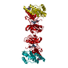
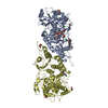
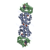
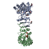
- PDB-2jc6: Crystal structure of human calmodulin-dependent protein kinase 1D -

+
Open data


ID or keywords:

Loading...

-
Basic information

Entry
Database: PDB / ID: 2jc6
TitleCrystal structure of human calmodulin-dependent protein kinase 1D
ComponentsCALCIUM/CALMODULIN-DEPENDENT PROTEIN KINASE TYPE 1D
KeywordsTRANSFERASE / ATP-BINDING / NUCLEAR PROTEIN / PHOSPHORYLATION
Function / homology
Function and homology information


regulation of granulocyte chemotaxis / positive regulation of CREB transcription factor activity / regulation of dendrite development / Ca2+/calmodulin-dependent protein kinase / positive regulation of neutrophil chemotaxis / calmodulin-dependent protein kinase activity / positive regulation of respiratory burst / positive regulation of phagocytosis / positive regulation of neuron projection development / nervous system development ...regulation of granulocyte chemotaxis / positive regulation of CREB transcription factor activity / regulation of dendrite development / Ca2+/calmodulin-dependent protein kinase / positive regulation of neutrophil chemotaxis / calmodulin-dependent protein kinase activity / positive regulation of respiratory burst / positive regulation of phagocytosis / positive regulation of neuron projection development / nervous system development / calmodulin binding / inflammatory response / positive regulation of apoptotic process / phosphorylation / protein serine kinase activity / negative regulation of apoptotic process / ATP binding / nucleus / cytoplasm
Similarity search - Function
Transferase(Phosphotransferase) domain 1 / Transferase(Phosphotransferase); domain 1 / Phosphorylase Kinase; domain 1 / Phosphorylase Kinase; domain 1 / Serine/threonine-protein kinase, active site / Serine/Threonine protein kinases active-site signature. / Protein kinase domain / Serine/Threonine protein kinases, catalytic domain / Protein kinase, ATP binding site / Protein kinases ATP-binding region signature. ...Transferase(Phosphotransferase) domain 1 / Transferase(Phosphotransferase); domain 1 / Phosphorylase Kinase; domain 1 / Phosphorylase Kinase; domain 1 / Serine/threonine-protein kinase, active site / Serine/Threonine protein kinases active-site signature. / Protein kinase domain / Serine/Threonine protein kinases, catalytic domain / Protein kinase, ATP binding site / Protein kinases ATP-binding region signature. / Protein kinase domain profile. / Protein kinase domain / Protein kinase-like domain superfamily / 2-Layer Sandwich / Orthogonal Bundle / Mainly Alpha / Alpha Beta
Similarity search - Domain/homology
Chem-QPP / Calcium/calmodulin-dependent protein kinase type 1D
Similarity search - Component
Biological speciesHOMO SAPIENS (human)
MethodX-RAY DIFFRACTION / SYNCHROTRON / MOLECULAR REPLACEMENT / Resolution: 2.3 Å
AuthorsDebreczeni, J.E. / Rellos, P. / Fedorov, O. / Niesen, F.H. / Bhatia, C. / Shrestha, L. / Salah, E. / Smee, C. / Colebrook, S. / Berridge, G. ...Debreczeni, J.E. / Rellos, P. / Fedorov, O. / Niesen, F.H. / Bhatia, C. / Shrestha, L. / Salah, E. / Smee, C. / Colebrook, S. / Berridge, G. / Gileadi, O. / Bunkoczi, G. / Ugochukwu, E. / Pike, A.C.W. / von Delft, F. / Knapp, S. / Sundstrom, M. / Weigelt, J. / Arrowsmith, C.H. / Edwards, A.
CitationJournal: To be Published
Title: Crystal Structure of Human Calmodulin-Dependent Protein Kinase 1D
Authors: Debreczeni, J.E. / Rellos, P. / Fedorov, O. / Niesen, F.H. / Bhatia, C. / Shrestha, L. / Salah, E. / Smee, C. / Colebrook, S. / Berridge, G. / Gileadi, O. / Bunkoczi, G. / Ugochukwu, E. / ...Authors: Debreczeni, J.E. / Rellos, P. / Fedorov, O. / Niesen, F.H. / Bhatia, C. / Shrestha, L. / Salah, E. / Smee, C. / Colebrook, S. / Berridge, G. / Gileadi, O. / Bunkoczi, G. / Ugochukwu, E. / Pike, A.C.W. / von Delft, F. / Knapp, S. / Sundstrom, M. / Weigelt, J. / Arrowsmith, C.H. / Edwards, A.
History
DepositionDec 19, 2006Deposition site: PDBE / Processing site: PDBE
Revision 1.0Feb 13, 2007Provider: repository / Type: Initial release
Revision 1.1Jul 13, 2011Group: Refinement description / Version format compliance
Revision 1.2Jan 24, 2018Group: Database references / Category: citation_author / Item: _citation_author.name
Revision 1.3Dec 13, 2023Group: Data collection / Database references ...Data collection / Database references / Other / Refinement description
Category: chem_comp_atom / chem_comp_bond ...chem_comp_atom / chem_comp_bond / database_2 / pdbx_database_status / pdbx_initial_refinement_model
Item: _database_2.pdbx_DOI / _database_2.pdbx_database_accession / _pdbx_database_status.status_code_sf

-
Structure visualization

Structure viewerMolecule:
MolmilJmol/JSmol

Downloads & links

-
Assembly

Deposited unit
A: CALCIUM/CALMODULIN-DEPENDENT PROTEIN KINASE TYPE 1D
C: CALCIUM/CALMODULIN-DEPENDENT PROTEIN KINASE TYPE 1D
hetero molecules


Theoretical massNumber of molelcules
Total (without water)76,3834
Polymers75,7802
Non-polymers6032
Water3,387188
1


  • Idetical with deposited unit
  • defined by author&software
TypeNameSymmetry operationNumber
identity operation1_555x,y,z1
Buried area3430 Å2
ΔGint-21.4 kcal/mol
Surface area27780 Å2
MethodPISA
Unit cell
Length a, b, c (Å)39.640, 93.060, 101.860
Angle α, β, γ (deg.)90.00, 96.16, 90.00
Int Tables number4
Space group name H-MP1211
Noncrystallographic symmetry (NCS)NCS domain:
IDEns-IDDetails
11A
21C

NCS domain segments:
Dom-IDComponent-IDEns-IDRefine codeAuth asym-IDAuth seq-ID
1114A3 - 222
2114C3 - 222
1214A233 - 313
2214C233 - 313

NCS oper: (Code: given
Matrix: (-0.99999, 0.00435, 0.00247), (0.00467, -0.63478, -0.77268), (-0.00179, -0.77268, 0.63479)
Vector: -4.30173, -126.35943, -59.53677)

-
Components

#1: Protein CALCIUM/CALMODULIN-DEPENDENT PROTEIN KINASE TYPE 1D / CALMODULIN-DEPENDENT PROTEIN KINASE 1D / CAM KINASE ID / CAM KINASE I DELTA / CAMKI-DELTA / CAM-KI ...CALMODULIN-DEPENDENT PROTEIN KINASE 1D / CAM KINASE ID / CAM KINASE I DELTA / CAMKI-DELTA / CAM-KI DELTA / CAMKI DELTA / CAMK1D / CAMKI-LIKE PROTEIN KINASE / CKLIK


Mass: 37890.125 Da / Num. of mol.: 2 / Fragment: RESIDUES 1-333
Source method: isolated from a genetically manipulated source
Source: (gene. exp.) HOMO SAPIENS (human) / Production host: ESCHERICHIA COLI (E. coli) / Strain (production host): BL21(DE3) / Variant (production host): R3-ROSETTA
References: UniProt: Q8IU85, Ca2+/calmodulin-dependent protein kinase
#2: Chemical ChemComp-QPP / N-(5-METHYL-1H-PYRAZOL-3-YL)-2-PHENYLQUINAZOLIN-4-AMINE / 5,6-DIHYDRO-BENZO[H]CINNOLIN-3-YLAMINE


Mass: 301.345 Da / Num. of mol.: 2 / Source method: obtained synthetically / Formula: C18H15N5
#3: Water ChemComp-HOH / water / Water


Mass: 18.015 Da / Num. of mol.: 188 / Source method: isolated from a natural source / Formula: H2O

-
Experimental details

-
Experiment

ExperimentMethod: X-RAY DIFFRACTION / Number of used crystals: 1

-
Sample preparation

CrystalDensity Matthews: 2.59 Å3/Da / Density % sol: 52 %
Crystal growDetails: 0.1M CITRATE PH 5.6, 20% ISOPROPANOL, 20% PEG 4K.

-
Data collection

DiffractionMean temperature: 100 K
Diffraction sourceSource: SYNCHROTRON / Site: SLS / Beamline: X10SA / Wavelength: 0.9796
DetectorType: MARRESEARCH / Detector: CCD / Date: Nov 27, 2006 / Details: MIRROR
RadiationMonochromator: SI 1 1 1 / Protocol: SINGLE WAVELENGTH / Monochromatic (M) / Laue (L): M / Scattering type: x-ray
Radiation wavelengthWavelength: 0.9796 Å / Relative weight: 1
ReflectionResolution: 2.3→42.2 Å / Num. obs: 29986 / % possible obs: 91.6 % / Observed criterion σ(I): 2 / Redundancy: 3.47 % / Rmerge(I) obs: 0.1 / Net I/σ(I): 11.17
Reflection shellResolution: 2.3→2.4 Å / Redundancy: 3.8 % / Rmerge(I) obs: 0.44 / Mean I/σ(I) obs: 2.56 / % possible all: 99.9

-
Processing

Software
NameVersionClassification
REFMAC5.2.0019refinement
XDSdata reduction
XDSdata scaling
PHASERphasing
RefinementMethod to determine structure: MOLECULAR REPLACEMENT
Starting model: PDB ENTRY 1A06
Resolution: 2.3→101.02 Å / Cor.coef. Fo:Fc: 0.941 / Cor.coef. Fo:Fc free: 0.898 / SU B: 13.019 / SU ML: 0.164 / TLS residual ADP flag: UNVERIFIED / Cross valid method: THROUGHOUT / ESU R: 0.306 / ESU R Free: 0.241 / Stereochemistry target values: MAXIMUM LIKELIHOOD / Details: HYDROGENS HAVE BEEN ADDED IN THE RIDING POSITIONS.
RfactorNum. reflection% reflectionSelection details
Rfree0.248 1514 5.1 %RANDOM
Rwork0.186 ---
obs0.189 28455 91.6 %-
Solvent computationIon probe radii: 0.8 Å / Shrinkage radii: 0.8 Å / VDW probe radii: 1.4 Å / Solvent model: MASK
Displacement parametersBiso mean: 11.63 Å2
Baniso -1Baniso -2Baniso -3
1--0.79 Å20 Å2-0.23 Å2
2--0.12 Å20 Å2
3---0.63 Å2
Refinement stepCycle: LAST / Resolution: 2.3→101.02 Å
ProteinNucleic acidLigandSolventTotal
Num. atoms4367 0 46 188 4601
Refine LS restraints
Refine-IDTypeDev idealDev ideal targetNumber
X-RAY DIFFRACTIONr_bond_refined_d0.0130.0224549
X-RAY DIFFRACTIONr_bond_other_d0.0040.023044
X-RAY DIFFRACTIONr_angle_refined_deg1.4281.9656169
X-RAY DIFFRACTIONr_angle_other_deg0.9173.0017399
X-RAY DIFFRACTIONr_dihedral_angle_1_deg9.2915555
X-RAY DIFFRACTIONr_dihedral_angle_2_deg39.5424.519208
X-RAY DIFFRACTIONr_dihedral_angle_3_deg17.51915754
X-RAY DIFFRACTIONr_dihedral_angle_4_deg23.3451520
X-RAY DIFFRACTIONr_chiral_restr0.0850.2667
X-RAY DIFFRACTIONr_gen_planes_refined0.0050.025064
X-RAY DIFFRACTIONr_gen_planes_other0.0010.02920
X-RAY DIFFRACTIONr_nbd_refined0.1890.2848
X-RAY DIFFRACTIONr_nbd_other0.190.23012
X-RAY DIFFRACTIONr_nbtor_refined0.180.22158
X-RAY DIFFRACTIONr_nbtor_other0.0890.22360
X-RAY DIFFRACTIONr_xyhbond_nbd_refined0.1730.2153
X-RAY DIFFRACTIONr_xyhbond_nbd_other
X-RAY DIFFRACTIONr_metal_ion_refined
X-RAY DIFFRACTIONr_metal_ion_other
X-RAY DIFFRACTIONr_symmetry_vdw_refined0.1060.213
X-RAY DIFFRACTIONr_symmetry_vdw_other0.2180.243
X-RAY DIFFRACTIONr_symmetry_hbond_refined0.1990.216
X-RAY DIFFRACTIONr_symmetry_hbond_other
X-RAY DIFFRACTIONr_symmetry_metal_ion_refined
X-RAY DIFFRACTIONr_symmetry_metal_ion_other
X-RAY DIFFRACTIONr_mcbond_it3.18232906
X-RAY DIFFRACTIONr_mcbond_other
X-RAY DIFFRACTIONr_mcangle_it4.33954446
X-RAY DIFFRACTIONr_mcangle_other
X-RAY DIFFRACTIONr_scbond_it6.69772014
X-RAY DIFFRACTIONr_scbond_other
X-RAY DIFFRACTIONr_scangle_it8.199111720
X-RAY DIFFRACTIONr_scangle_other
X-RAY DIFFRACTIONr_long_range_B_refined
X-RAY DIFFRACTIONr_long_range_B_other
X-RAY DIFFRACTIONr_rigid_bond_restr
X-RAY DIFFRACTIONr_sphericity_free
X-RAY DIFFRACTIONr_sphericity_bonded
Refine LS restraints NCS

Dom-ID: 1 / Auth asym-ID: A / Ens-ID: 1 / Number: 3518 / Refine-ID: X-RAY DIFFRACTION

TypeRms dev position (Å)Weight position
medium positional0.250.5
medium thermal0.922
LS refinement shellResolution: 2.3→2.36 Å / Total num. of bins used: 20 /
RfactorNum. reflection
Rfree0.287 117
Rwork0.213 2276
Refinement TLS params.

Method: refined / Refine-ID: X-RAY DIFFRACTION

IDL112)L122)L132)L222)L232)L332)S11 (Å °)S12 (Å °)S13 (Å °)S21 (Å °)S22 (Å °)S23 (Å °)S31 (Å °)S32 (Å °)S33 (Å °)T112)T122)T132)T222)T232)T332)Origin x (Å)Origin y (Å)Origin z (Å)
12.94362.20910.7964.55353.1373.06230.2721-0.43930.33270.3328-0.28170.21870.065-0.09170.0096-0.0407-0.0858-0.0001-0.0604-0.03580.0132-8.105-74.783-29.44
21.8026-0.155-0.04721.19840.15421.17090.1037-0.0897-0.0454-0.0537-0.05070.0150.08930.0229-0.053-0.1105-0.084-0.005-0.0597-0.0147-0.1081.907-97.572-44.083
31.6125-0.5763-0.30413.77220.24311.29210.0726-0.0783-0.07850.2252-0.06240.09020.2036-0.0051-0.0102-0.0498-0.0744-0.0237-0.0622-0.0115-0.10893.643-56.113-20.569
42.86770.1224-0.07461.37410.3921.11040.0978-0.21210.347-0.0035-0.0430.0224-0.15850.02-0.0548-0.0835-0.087-0.0023-0.0819-0.0321-0.0237-6.763-30.25-12.18
Refinement TLS group
IDRefine-IDRefine TLS-IDAuth asym-IDAuth seq-ID
1X-RAY DIFFRACTION1A11 - 101
2X-RAY DIFFRACTION2A102 - 313
3X-RAY DIFFRACTION3C11 - 101
4X-RAY DIFFRACTION4C102 - 313

+
About Yorodumi

-
News

-
Feb 9, 2022. New format data for meta-information of EMDB entries

New format data for meta-information of EMDB entries

  • Version 3 of the EMDB header file is now the official format.
  • The previous official version 1.9 will be removed from the archive.

Related info.:EMDB header

External links:wwPDB to switch to version 3 of the EMDB data model

-
Aug 12, 2020. Covid-19 info

Covid-19 info

URL: https://pdbj.org/emnavi/covid19.php

New page: Covid-19 featured information page in EM Navigator.

Related info.:Covid-19 info / Mar 5, 2020. Novel coronavirus structure data

+
Mar 5, 2020. Novel coronavirus structure data

Novel coronavirus structure data

Related info.:Yorodumi Speices / Aug 12, 2020. Covid-19 info

External links:COVID-19 featured content - PDBj / Molecule of the Month (242):Coronavirus Proteases

+
Jan 31, 2019. EMDB accession codes are about to change! (news from PDBe EMDB page)

EMDB accession codes are about to change! (news from PDBe EMDB page)

  • The allocation of 4 digits for EMDB accession codes will soon come to an end. Whilst these codes will remain in use, new EMDB accession codes will include an additional digit and will expand incrementally as the available range of codes is exhausted. The current 4-digit format prefixed with “EMD-” (i.e. EMD-XXXX) will advance to a 5-digit format (i.e. EMD-XXXXX), and so on. It is currently estimated that the 4-digit codes will be depleted around Spring 2019, at which point the 5-digit format will come into force.
  • The EM Navigator/Yorodumi systems omit the EMD- prefix.

Related info.:Q: What is EMD? / ID/Accession-code notation in Yorodumi/EM Navigator

External links:EMDB Accession Codes are Changing Soon! / Contact to PDBj

+
Jul 12, 2017. Major update of PDB

Major update of PDB

  • wwPDB released updated PDB data conforming to the new PDBx/mmCIF dictionary.
  • This is a major update changing the version number from 4 to 5, and with Remediation, in which all the entries are updated.
  • In this update, many items about electron microscopy experimental information are reorganized (e.g. em_software).
  • Now, EM Navigator and Yorodumi are based on the updated data.

External links:wwPDB Remediation / Enriched Model Files Conforming to OneDep Data Standards Now Available in the PDB FTP Archive

-
Yorodumi

Thousand views of thousand structures

  • Yorodumi is a browser for structure data from EMDB, PDB, SASBDB, etc.
  • This page is also the successor to EM Navigator detail page, and also detail information page/front-end page for Omokage search.
  • The word "yorodu" (or yorozu) is an old Japanese word meaning "ten thousand". "mi" (miru) is to see.

Related info.:EMDB / PDB / SASBDB / Comparison of 3 databanks / Yorodumi Search / Aug 31, 2016. New EM Navigator & Yorodumi / Yorodumi Papers / Jmol/JSmol / Function and homology information / Changes in new EM Navigator and Yorodumi

Read more