- PDB-2j3r: The crystal structure of the bet3-trs31 heterodimer. -
+
Open data
ID or keywords:
Loading...
-
Basic information
Entry
Database: PDB / ID: 2j3r
Title
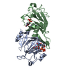
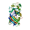
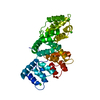
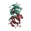
The crystal structure of the bet3-trs31 heterodimer.
Components
TRAFFICKING PROTEIN PARTICLE COMPLEX SUBUNIT 3
ZGC 92866
Keywords
TRANSPORT / GOLGI APPARATUS / VESICLE TRANSPORT / ER-GOLGI TRANSPORT / TRAPP / PALMITATE / LIPOPROTEIN / ENDOPLASMIC RETICULUM / MULTISUBUNIT TETHERING FACTOR
Function / homology
Function and homology information
COPII-mediated vesicle transport / vesicle coating / vesicle tethering / RAB GEFs exchange GTP for GDP on RABs / TRAPPI protein complex / COPII-mediated vesicle transport / TRAPPII protein complex / TRAPPIII protein complex / TRAPP complex / COPII vesicle coating ...COPII-mediated vesicle transport / vesicle coating / vesicle tethering / RAB GEFs exchange GTP for GDP on RABs / TRAPPI protein complex / COPII-mediated vesicle transport / TRAPPII protein complex / TRAPPIII protein complex / TRAPP complex / COPII vesicle coating / endoplasmic reticulum to Golgi vesicle-mediated transport / Golgi membrane / endoplasmic reticulum / cytoplasm / cytosol Similarity search - Function
Trafficking protein particle complex subunit 3 / TRAPP I complex, subunit 5 / Bet3 family / Transport protein particle (TRAPP) component / Transport protein particle (TRAPP) component / Muramoyl-pentapeptide Carboxypeptidase; domain 2 / NO signalling/Golgi transport ligand-binding domain superfamily / 2-Layer Sandwich / Alpha Beta Similarity search - Domain/homology
NITRATE ION / PALMITIC ACID / Trafficking protein particle complex subunit 3 / Trafficking protein particle complex subunit 5 Similarity search - Component
In the structure databanks used in Yorodumi, some data are registered as the other names, "COVID-19 virus" and "2019-nCoV". Here are the details of the virus and the list of structure data.
Jan 31, 2019. EMDB accession codes are about to change! (news from PDBe EMDB page)
EMDB accession codes are about to change! (news from PDBe EMDB page)
The allocation of 4 digits for EMDB accession codes will soon come to an end. Whilst these codes will remain in use, new EMDB accession codes will include an additional digit and will expand incrementally as the available range of codes is exhausted. The current 4-digit format prefixed with “EMD-” (i.e. EMD-XXXX) will advance to a 5-digit format (i.e. EMD-XXXXX), and so on. It is currently estimated that the 4-digit codes will be depleted around Spring 2019, at which point the 5-digit format will come into force.
The EM Navigator/Yorodumi systems omit the EMD- prefix.
Related info.:Q: What is EMD? / ID/Accession-code notation in Yorodumi/EM Navigator
Yorodumi is a browser for structure data from EMDB, PDB, SASBDB, etc.
This page is also the successor to EM Navigator detail page, and also detail information page/front-end page for Omokage search.
The word "yorodu" (or yorozu) is an old Japanese word meaning "ten thousand". "mi" (miru) is to see.
Related info.:EMDB / PDB / SASBDB / Comparison of 3 databanks / Yorodumi Search / Aug 31, 2016. New EM Navigator & Yorodumi / Yorodumi Papers / Jmol/JSmol / Function and homology information / Changes in new EM Navigator and Yorodumi
 Movie
Movie Controller


 Open data
 Basic information
 Components
 Keywords
 Function and homology information

 X-RAY DIFFRACTION /
  Authors
 Citation
 Structure visualization
 Downloads & links
 Other downloads


 PDBj
PDBj


 Assembly


 Mass: 256.424 Da / Num. of mol.: 1 / Source method: obtained synthetically / Formula: C16H32O2
 Mass: 62.005 Da / Num. of mol.: 1 / Source method: obtained synthetically / Formula: NO3
 Mass: 18.015 Da / Num. of mol.: 31 / Source method: isolated from a natural source / Formula: H2O
 Sample preparation
 / Beamline: AR-NW12A / Wavelength: 1
  Processing