 Movie
Movie Controller

[English] 日本語
 Yorodumi
- PDB-1axn: THE HIGH RESOLUTION STRUCTURE OF ANNEXIN III SHOWS DIFFERENCES WI... -
+ Open data

- Basic information
| Entry | Database: PDB / ID: 1axn | ||||||
|---|---|---|---|---|---|---|---|
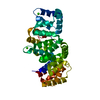
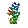
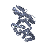
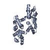
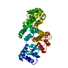
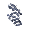
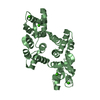
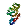
| Title | THE HIGH RESOLUTION STRUCTURE OF ANNEXIN III SHOWS DIFFERENCES WITH ANNEXIN V | ||||||
 Components Components | ANNEXIN III | ||||||
 Keywords Keywords | CALCIUM/PHOSPHOLIPID-BINDING PROTEIN / ANNEXIN FAMILY / CALCIUM-PHOSPHOLIPID-BINDING PROTEIN COMPLEX | ||||||
| Function / homology |  Function and homology information Function and homology informationneutrophil degranulation / phospholipase A2 inhibitor activity / specific granule / calcium-dependent phospholipid binding / vesicle membrane / phosphatidylserine binding / : / phagocytosis / positive regulation of endothelial cell migration / Developmental Lineage of Pancreatic Ductal Cells ...neutrophil degranulation / phospholipase A2 inhibitor activity / specific granule / calcium-dependent phospholipid binding / vesicle membrane / phosphatidylserine binding / : / phagocytosis / positive regulation of endothelial cell migration / Developmental Lineage of Pancreatic Ductal Cells / phagocytic vesicle membrane / positive regulation of angiogenesis / calcium-dependent protein binding / defense response to bacterium / calcium ion binding / extracellular exosome / membrane / nucleus / plasma membrane / cytoplasm Similarity search - Function | ||||||
| Biological species |  Homo sapiens (human) Homo sapiens (human) | ||||||
| Method |  X-RAY DIFFRACTION / SYNCHROTRON / Resolution: 1.78 Å X-RAY DIFFRACTION / SYNCHROTRON / Resolution: 1.78 Å | ||||||
 Authors Authors | Favier-Perron, B. / Lewit-Bentley, A. / Russo-Marie, F. | ||||||
 Citation Citation | Journal: Biochemistry / Year: 1996 Title: The high-resolution crystal structure of human annexin III shows subtle differences with annexin V. Authors: Favier-Perron, B. / Lewit-Bentley, A. / Russo-Marie, F. | ||||||
| History |
|
- Structure visualization
| Structure viewer | Molecule: MolmilJmol/JSmol |
|---|
- Downloads & links
-Download
| PDBx/mmCIF format | 1axn.cif.gz | 82.7 KB | Display | PDBx/mmCIF format |
|---|---|---|---|---|
| PDB format | pdb1axn.ent.gz | 61.5 KB | Display | PDB format |
| PDBx/mmJSON format | 1axn.json.gz | Tree view | PDBx/mmJSON format | |
| Others |  Other downloads Other downloads |
-Validation report
| Arichive directory | https://data.pdbj.org/pub/pdb/validation_reports/ax/1axnftp://data.pdbj.org/pub/pdb/validation_reports/ax/1axn | HTTPS FTP |
|---|
-Related structure data
| Similar structure data |
|---|


-Links
 PDBj
PDBj- Assembly
| Deposited unit | 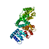
| ||||||||
|---|---|---|---|---|---|---|---|---|---|
| 1 |
| ||||||||
| Unit cell |
|
-Components
| #1: Protein | Mass: 36383.148 Da / Num. of mol.: 1 Source method: isolated from a genetically manipulated source Details: HUMAN RECOMBINANT / Source: (gene. exp.) Homo sapiens (human) / Description: PREPARED AS A GST-FUSION, THEN CLEAVED YES / Gene: HUMAN ANNEXIN III / Plasmid: PGEX2T / Gene (production host): HUMAN ANNEXIN III / Production host:  | ||
|---|---|---|---|
| #2: Chemical | ChemComp-CA /   Mass: 40.078 Da / Num. of mol.: 5 / Source method: obtained synthetically / Formula: Ca Mass: 40.078 Da / Num. of mol.: 5 / Source method: obtained synthetically / Formula: Ca#3: Water | ChemComp-HOH / |  Mass: 18.015 Da / Num. of mol.: 281 / Source method: isolated from a natural source / Formula: H2O Mass: 18.015 Da / Num. of mol.: 281 / Source method: isolated from a natural source / Formula: H2O |
-Experimental details
-Experiment
| Experiment | Method: X-RAY DIFFRACTION |
|---|
- Sample preparation
| Crystal | Density Matthews: 2.05 Å3/Da / Density % sol: 39.94 % | ||||||||||||||||||||||||||||||||||||
|---|---|---|---|---|---|---|---|---|---|---|---|---|---|---|---|---|---|---|---|---|---|---|---|---|---|---|---|---|---|---|---|---|---|---|---|---|---|
| Crystal | *PLUS Density % sol: 42 % | ||||||||||||||||||||||||||||||||||||
| Crystal grow | *PLUS pH: 7.5 / Method: vapor diffusion, hanging drop | ||||||||||||||||||||||||||||||||||||
| Components of the solutions | *PLUS
|
-Data collection
| Diffraction source | Source: SYNCHROTRON / Site: LURE  / Beamline: DW32 / Wavelength: 0.9 / Beamline: DW32 / Wavelength: 0.9 |
|---|---|
| Detector | Type: MARRESEARCH / Detector: IMAGE PLATE / Date: Apr 13, 1994 |
| Radiation | Monochromatic (M) / Laue (L): M / Scattering type: x-ray |
| Radiation wavelength | Wavelength: 0.9 Å / Relative weight: 1 |
| Reflection | Resolution: 1.78→100 Å / Num. obs: 25718 / % possible obs: 92 % / Observed criterion σ(I): 0 / Redundancy: 3.45 % / Rmerge(I) obs: 0.043 |
| Reflection | *PLUS Lowest resolution: 23.8 Å / Num. measured all: 88603 / Rmerge(I) obs: 0.043 |
| Reflection shell | *PLUS Highest resolution: 1.78 Å / Lowest resolution: 1.83 Å / % possible obs: 79.2 % |
- Processing
| Software |
| ||||||||||||||||||||||||||||||||||||||||||||||||||||||||||||||||||||||||||||||||||||
|---|---|---|---|---|---|---|---|---|---|---|---|---|---|---|---|---|---|---|---|---|---|---|---|---|---|---|---|---|---|---|---|---|---|---|---|---|---|---|---|---|---|---|---|---|---|---|---|---|---|---|---|---|---|---|---|---|---|---|---|---|---|---|---|---|---|---|---|---|---|---|---|---|---|---|---|---|---|---|---|---|---|---|---|---|---|
| Refinement | Resolution: 1.78→8 Å / σ(F): 2 /
| ||||||||||||||||||||||||||||||||||||||||||||||||||||||||||||||||||||||||||||||||||||
| Displacement parameters | Biso mean: 20.2 Å2 | ||||||||||||||||||||||||||||||||||||||||||||||||||||||||||||||||||||||||||||||||||||
| Refinement step | Cycle: LAST / Resolution: 1.78→8 Å
| ||||||||||||||||||||||||||||||||||||||||||||||||||||||||||||||||||||||||||||||||||||
| Refine LS restraints |
| ||||||||||||||||||||||||||||||||||||||||||||||||||||||||||||||||||||||||||||||||||||
| Software | *PLUS Name: PROLSQ / Classification: refinement | ||||||||||||||||||||||||||||||||||||||||||||||||||||||||||||||||||||||||||||||||||||
| Refinement | *PLUS Rfactor obs: 0.173 / Rfactor Rfree: 0.242 | ||||||||||||||||||||||||||||||||||||||||||||||||||||||||||||||||||||||||||||||||||||
| Solvent computation | *PLUS | ||||||||||||||||||||||||||||||||||||||||||||||||||||||||||||||||||||||||||||||||||||
| Displacement parameters | *PLUS | ||||||||||||||||||||||||||||||||||||||||||||||||||||||||||||||||||||||||||||||||||||
| Refine LS restraints | *PLUS
|
