- PDB-2i02: CRYSTAL STRUCTURE OF a pyridoxamine 5'-phosphate oxidase-like fam... -
+
Open data
ID or keywords:
Loading...
-
Basic information
Entry
Database: PDB / ID: 2i02
Title
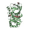
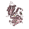
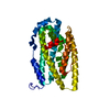
CRYSTAL STRUCTURE OF a pyridoxamine 5'-phosphate oxidase-like family protein (NPUN_R6570) FROM NOSTOC PUNCTIFORME PCC 73102 AT 1.80 A RESOLUTION
Components
general stress protein of COG3871
Keywords
OXIDOREDUCTASE / GENERAL STRESS PROTEIN OF COG3871 / PYRIDOXAMINE 5'-PHOSPHATE OXIDASE-LIKE FAMILY PROTEIN / STRUCTURAL GENOMICS / JOINT CENTER FOR STRUCTURAL GENOMICS / JCSG / PROTEIN STRUCTURE INITIATIVE / PSI-2
Monochromator: Single crystal Si(111) bent monochromator (horizontal focusing) Protocol: MAD / Monochromatic (M) / Laue (L): M / Scattering type: x-ray
Radiation wavelength
ID
Wavelength (Å)
Relative weight
1
0.979197
1
2
0.91837
1
Reflection
Resolution: 1.8→29.921 Å / Num. obs: 32823 / % possible obs: 100 % / Redundancy: 10.3 % / Rmerge(I) obs: 0.117 / Rsym value: 0.117 / Net I/σ(I): 4.8
Reflection shell
Diffraction-ID: 1
Resolution (Å)
Redundancy (%)
Rmerge(I) obs
Mean I/σ(I) obs
Num. measured all
Num. unique all
Rsym value
% possible all
1.8-1.85
10.6
0.946
0.8
25296
2377
0.946
100
1.85-1.9
10.6
0.731
1.1
24511
2311
0.731
100
1.9-1.95
10.6
0.543
1.4
24005
2262
0.543
100
1.95-2.01
10.6
0.435
1.8
23213
2187
0.435
100
2.01-2.08
10.6
0.36
2.1
22570
2136
0.36
100
2.08-2.15
10.6
0.303
2.5
21862
2068
0.303
100
2.15-2.23
10.6
0.255
3
20934
1983
0.255
100
2.23-2.32
10.5
0.205
3.7
20335
1938
0.205
100
2.32-2.43
10.4
0.181
4.2
19221
1842
0.181
100
2.43-2.55
10.5
0.161
4.7
18552
1772
0.161
100
2.55-2.68
10.4
0.134
5.5
17591
1694
0.134
100
2.68-2.85
10.3
0.113
6.5
16658
1610
0.113
100
2.85-3.04
10.3
0.086
8.1
15668
1527
0.086
100
3.04-3.29
10.2
0.079
8.2
14363
1413
0.079
100
3.29-3.6
10
0.075
8.2
13195
1314
0.075
100
3.6-4.02
9.7
0.067
8.9
11638
1200
0.067
100
4.02-4.65
9
0.07
8.3
9675
1074
0.07
100
4.65-5.69
9.7
0.065
8.9
8980
925
0.065
100
5.69-8.05
9.9
0.057
11.2
7313
741
0.057
100
8.05-29.92
8.5
0.045
12.3
3829
449
0.045
98.2
-
Phasing
Phasing
Method: MAD
-
Processing
Software
Name
Version
Classification
NB
MolProbity
3beta29
modelbuilding
SHELX
phasing
REFMAC
5.2.0019
refinement
SCALA
datascaling
PDB_EXTRACT
2
dataextraction
MOSFLM
datareduction
CCP4
(SCALA)
datascaling
SHELXD
phasing
SHARP
phasing
Refinement
Method to determine structure: MAD / Resolution: 1.8→29.921 Å / Cor.coef. Fo:Fc: 0.959 / Cor.coef. Fo:Fc free: 0.95 / SU B: 4.503 / SU ML: 0.071 / TLS residual ADP flag: LIKELY RESIDUAL / Cross valid method: THROUGHOUT / σ(F): 0 / ESU R: 0.113 / ESU R Free: 0.107 Stereochemistry target values: MAXIMUM LIKELIHOOD WITH PHASES Details: 1. HYDROGENS HAVE BEEN ADDED IN THE RIDING POSITIONS. 2. A MET-INHIBITION PROTOCOL WAS USED FOR SELENOMETHIONINE INCORPORATION DURING PROTEIN EXPRESSION. THE OCCUPANCY OF THE SE ATOMS IN THE ...Details: 1. HYDROGENS HAVE BEEN ADDED IN THE RIDING POSITIONS. 2. A MET-INHIBITION PROTOCOL WAS USED FOR SELENOMETHIONINE INCORPORATION DURING PROTEIN EXPRESSION. THE OCCUPANCY OF THE SE ATOMS IN THE MSE RESIDUES WAS REDUCED TO 0.75 FOR THE REDUCED SCATTERING POWER DUE TO PARTIAL S-MET INCORPORATION. 3. RESIDUES A1-4, A46-48, B1-4, AND B136-138 ARE DISORDERED AND NOT INCLUDED IN THE MODEL. 4. RESIDUES B45-48 AND B61 ARE POORLY ORDERED. 5. MOLECULES FMN AND PEG-300 (P33) ARE MODELED. 6. ATOM RECORDS CONTAIN RESIDUAL B FACTORS ONLY. 7. UNEXPLAINED DENSITY OBSERVED NEAR RESIDUES A58, A98, A101, B31-32, B61, B67.
Rfactor
Num. reflection
% reflection
Selection details
Rfree
0.204
1663
5.1 %
RANDOM
Rwork
0.177
-
-
-
all
0.178
-
-
-
obs
-
32793
99.99 %
-
Solvent computation
Ion probe radii: 0.8 Å / Shrinkage radii: 0.8 Å / VDW probe radii: 1.2 Å / Solvent model: BABINET MODEL WITH MASK
Displacement parameters
Biso mean: 25.749 Å2
Baniso -1
Baniso -2
Baniso -3
1-
-0.55 Å2
-0.27 Å2
0 Å2
2-
-
-0.55 Å2
0 Å2
3-
-
-
0.82 Å2
Refinement step
Cycle: LAST / Resolution: 1.8→29.921 Å
Protein
Nucleic acid
Ligand
Solvent
Total
Num. atoms
2262
0
53
175
2490
Refine LS restraints
Refine-ID
Type
Dev ideal
Dev ideal target
Number
X-RAY DIFFRACTION
r_bond_refined_d
0.014
0.022
2445
X-RAY DIFFRACTION
r_bond_other_d
0.001
0.02
1633
X-RAY DIFFRACTION
r_angle_refined_deg
1.496
1.954
3329
X-RAY DIFFRACTION
r_angle_other_deg
0.88
3
4014
X-RAY DIFFRACTION
r_dihedral_angle_1_deg
6.242
5
294
X-RAY DIFFRACTION
r_dihedral_angle_2_deg
38.584
25.128
117
X-RAY DIFFRACTION
r_dihedral_angle_3_deg
13.209
15
418
X-RAY DIFFRACTION
r_dihedral_angle_4_deg
13.334
15
7
X-RAY DIFFRACTION
r_chiral_restr
0.085
0.2
362
X-RAY DIFFRACTION
r_gen_planes_refined
0.006
0.02
2657
X-RAY DIFFRACTION
r_gen_planes_other
0.002
0.02
473
X-RAY DIFFRACTION
r_nbd_refined
0.22
0.2
454
X-RAY DIFFRACTION
r_nbd_other
0.199
0.2
1661
X-RAY DIFFRACTION
r_nbtor_refined
0.185
0.2
1157
X-RAY DIFFRACTION
r_nbtor_other
0.088
0.2
1272
X-RAY DIFFRACTION
r_xyhbond_nbd_refined
0.158
0.2
148
X-RAY DIFFRACTION
r_symmetry_vdw_refined
0.188
0.2
29
X-RAY DIFFRACTION
r_symmetry_vdw_other
0.22
0.2
60
X-RAY DIFFRACTION
r_symmetry_hbond_refined
0.169
0.2
21
X-RAY DIFFRACTION
r_mcbond_it
2.36
3
1490
X-RAY DIFFRACTION
r_mcbond_other
0.568
3
567
X-RAY DIFFRACTION
r_mcangle_it
3.393
5
2344
X-RAY DIFFRACTION
r_scbond_it
5.215
8
1164
X-RAY DIFFRACTION
r_scangle_it
7.245
11
976
LS refinement shell
Resolution: 1.8→1.847 Å / Total num. of bins used: 20
Rfactor
Num. reflection
% reflection
Rfree
0.261
107
-
Rwork
0.21
2258
-
obs
-
2365
100 %
Refinement TLS params.
Method: refined / Refine-ID: X-RAY DIFFRACTION
ID
L11 (°2)
L12 (°2)
L13 (°2)
L22 (°2)
L23 (°2)
L33 (°2)
S11 (Å °)
S12 (Å °)
S13 (Å °)
S21 (Å °)
S22 (Å °)
S23 (Å °)
S31 (Å °)
S32 (Å °)
S33 (Å °)
T11 (Å2)
T12 (Å2)
T13 (Å2)
T22 (Å2)
T23 (Å2)
T33 (Å2)
Origin x (Å)
Origin y (Å)
Origin z (Å)
1
1.2444
0.2008
0.5195
1.5427
0.6023
1.6124
0.018
0.251
0.13
-0.2164
0.0291
-0.1648
-0.1179
0.176
-0.0471
-0.0412
-0.0179
0.0245
-0.0126
0.0174
-0.0316
28.997
25.119
15.445
2
1.5109
0.2729
-0.0151
0.3117
0.0651
1.0702
0.0114
0.159
0.1698
-0.1101
-0.0034
0.0602
-0.1216
-0.0855
-0.008
-0.0358
0.0154
-0.0007
-0.0321
0.0134
-0.0318
5.987
25.867
16.087
Refinement TLS group
Refine-ID: X-RAY DIFFRACTION / Selection: ALL
ID
Refine TLS-ID
Auth asym-ID
Label asym-ID
Auth seq-ID
Label seq-ID
1
1
A
A
5 - 45
6 - 46
2
1
A
A
49 - 147
50 - 148
3
2
B
B
5 - 135
6 - 136
4
2
B
B
139 - 147
140 - 148
+
About Yorodumi
-
News
-
Feb 9, 2022. New format data for meta-information of EMDB entries
New format data for meta-information of EMDB entries
Version 3 of the EMDB header file is now the official format.
The previous official version 1.9 will be removed from the archive.
In the structure databanks used in Yorodumi, some data are registered as the other names, "COVID-19 virus" and "2019-nCoV". Here are the details of the virus and the list of structure data.
Jan 31, 2019. EMDB accession codes are about to change! (news from PDBe EMDB page)
EMDB accession codes are about to change! (news from PDBe EMDB page)
The allocation of 4 digits for EMDB accession codes will soon come to an end. Whilst these codes will remain in use, new EMDB accession codes will include an additional digit and will expand incrementally as the available range of codes is exhausted. The current 4-digit format prefixed with “EMD-” (i.e. EMD-XXXX) will advance to a 5-digit format (i.e. EMD-XXXXX), and so on. It is currently estimated that the 4-digit codes will be depleted around Spring 2019, at which point the 5-digit format will come into force.
The EM Navigator/Yorodumi systems omit the EMD- prefix.
Related info.:Q: What is EMD? / ID/Accession-code notation in Yorodumi/EM Navigator
Yorodumi is a browser for structure data from EMDB, PDB, SASBDB, etc.
This page is also the successor to EM Navigator detail page, and also detail information page/front-end page for Omokage search.
The word "yorodu" (or yorozu) is an old Japanese word meaning "ten thousand". "mi" (miru) is to see.
Related info.:EMDB / PDB / SASBDB / Comparison of 3 databanks / Yorodumi Search / Aug 31, 2016. New EM Navigator & Yorodumi / Yorodumi Papers / Jmol/JSmol / Function and homology information / Changes in new EM Navigator and Yorodumi
 Movie
Movie Controller

 Yorodumi
 Open data
 Basic information
 Components
 Keywords
 Function and homology information
 Nostoc punctiforme (bacteria)
 X-RAY DIFFRACTION /
  Authors
 Citation
 Structure visualization
 Downloads & links
 Other downloads


 PDBj
PDBj Assembly
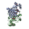

 Mass: 456.344 Da / Num. of mol.: 1 / Source method: obtained synthetically / Formula: C17H21N4O9P
 Mass: 326.383 Da / Num. of mol.: 2 / Source method: obtained synthetically / Formula: C14H30O8 / Comment: precipitant*YM
 Mass: 35.453 Da / Num. of mol.: 1 / Source method: obtained synthetically / Formula: Cl
 Mass: 18.015 Da / Num. of mol.: 175 / Source method: isolated from a natural source / Formula: H2O
 Sample preparation
 / Beamline: BL11-1 / Wavelength: 0.979197,0.918370
 Processing