Journal: Protein Sci. / Year: 2007 Title: Contributions of the interdomain loop, amino terminus, and subunit interface to the ligand-facilitated dimerization of neurophysin: crystal structures and mutation studies of bovine neurophysin-I. Authors: Li, X. / Lee, H. / Wu, J. / Breslow, E.
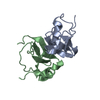
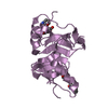
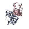
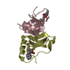
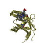
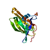
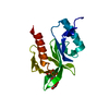
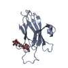
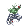
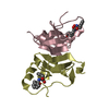
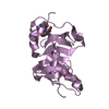
The biological unit is a dimer. There are 2.5 biological units per asymmetric unit. The complete dimers are comprised of chains A & B and chains C & D. Chain E is half of a dimer from another asymmetric unit.
-
Components
#1: Protein
Oxytocin-neurophysin1
Mass: 8134.256 Da / Num. of mol.: 5 / Fragment: Residues 38-118 / Mutation: Q58V Source method: isolated from a genetically manipulated source Source: (gene. exp.) Bos taurus (domestic cattle) / Gene: OXT / Plasmid: pTHMa30-51 / Production host: Escherichia coli (E. coli) / Strain (production host): BL21(DE3)p Lys S / References: UniProt: P01175
In the structure databanks used in Yorodumi, some data are registered as the other names, "COVID-19 virus" and "2019-nCoV". Here are the details of the virus and the list of structure data.
Jan 31, 2019. EMDB accession codes are about to change! (news from PDBe EMDB page)
EMDB accession codes are about to change! (news from PDBe EMDB page)
The allocation of 4 digits for EMDB accession codes will soon come to an end. Whilst these codes will remain in use, new EMDB accession codes will include an additional digit and will expand incrementally as the available range of codes is exhausted. The current 4-digit format prefixed with “EMD-” (i.e. EMD-XXXX) will advance to a 5-digit format (i.e. EMD-XXXXX), and so on. It is currently estimated that the 4-digit codes will be depleted around Spring 2019, at which point the 5-digit format will come into force.
The EM Navigator/Yorodumi systems omit the EMD- prefix.
Related info.:Q: What is EMD? / ID/Accession-code notation in Yorodumi/EM Navigator
Yorodumi is a browser for structure data from EMDB, PDB, SASBDB, etc.
This page is also the successor to EM Navigator detail page, and also detail information page/front-end page for Omokage search.
The word "yorodu" (or yorozu) is an old Japanese word meaning "ten thousand". "mi" (miru) is to see.
Related info.:EMDB / PDB / SASBDB / Comparison of 3 databanks / Yorodumi Search / Aug 31, 2016. New EM Navigator & Yorodumi / Yorodumi Papers / Jmol/JSmol / Function and homology information / Changes in new EM Navigator and Yorodumi
 Movie
Movie Controller

 Yorodumi
 Open data
 Basic information
 Components
 Keywords
 Function and homology information
 X-RAY DIFFRACTION / Resolution: 2.5 Å
  Authors
 Citation
 Structure visualization
 Downloads & links
 Other downloads
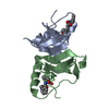


 PDBj
PDBj


 Assembly
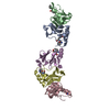


 Type: L-peptide linking / Mass: 165.189 Da / Num. of mol.: 5 / Source method: obtained synthetically / Formula: C9H11NO2
 Type: L-peptide linking / Mass: 181.189 Da / Num. of mol.: 5 / Source method: obtained synthetically / Formula: C9H11NO3
 Mass: 18.015 Da / Num. of mol.: 29 / Source method: isolated from a natural source / Formula: H2O
 Sample preparation
 Processing