 Movie
Movie Controller

[English] 日本語
 Yorodumi
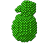
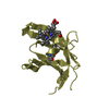
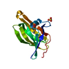
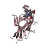
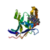
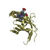
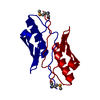
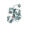
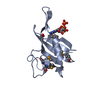
- PDB-5cvw: CRYSTAL STRUCTURE OF RTX DOMAIN BLOCK V OF ADENYLATE CYCLASE TOXI... -
+ Open data

- Basic information
| Entry | Database: PDB / ID: 5cvw | |||||||||
|---|---|---|---|---|---|---|---|---|---|---|
| Title | CRYSTAL STRUCTURE OF RTX DOMAIN BLOCK V OF ADENYLATE CYCLASE TOXIN FROM BORDETELLA PERTUSSIS | |||||||||
 Components Components | Bifunctional hemolysin/adenylate cyclase | |||||||||
 Keywords Keywords | TOXIN / ADENYLATE CYCLASE / RTX MOTIFS / CALCIUM BINDING | |||||||||
| Function / homology |  Function and homology information Function and homology informationcalcium- and calmodulin-responsive adenylate cyclase activity / hemolysis in another organism / host cell surface binding / adenylate cyclase / cAMP biosynthetic process / toxin activity / channel activity / calmodulin binding / calcium ion binding / host cell plasma membrane ...calcium- and calmodulin-responsive adenylate cyclase activity / hemolysis in another organism / host cell surface binding / adenylate cyclase / cAMP biosynthetic process / toxin activity / channel activity / calmodulin binding / calcium ion binding / host cell plasma membrane / extracellular region / ATP binding Similarity search - Function | |||||||||
| Biological species |  Bordetella pertussis (bacteria) Bordetella pertussis (bacteria) | |||||||||
| Method |  X-RAY DIFFRACTION / SYNCHROTRON / MOLECULAR REPLACEMENT / Resolution: 1.25 Å X-RAY DIFFRACTION / SYNCHROTRON / MOLECULAR REPLACEMENT / Resolution: 1.25 Å | |||||||||
 Authors Authors | Motlova, L. / Barinka, C. / Bumba, L. | |||||||||
| Funding support |  Czech Republic, 2items Czech Republic, 2items
| |||||||||
 Citation Citation | Journal: Mol Cell / Year: 2016 Title: Calcium-Driven Folding of RTX Domain β-Rolls Ratchets Translocation of RTX Proteins through Type I Secretion Ducts. Authors: Ladislav Bumba / Jiri Masin / Pavel Macek / Tomas Wald / Lucia Motlova / Ilona Bibova / Nela Klimova / Lucie Bednarova / Vaclav Veverka / Michael Kachala / Dmitri I Svergun / Cyril Barinka / Peter Sebo /  Abstract: Calcium-binding RTX proteins are equipped with C-terminal secretion signals and translocate from the Ca(2+)-depleted cytosol of Gram-negative bacteria directly into the Ca(2+)-rich external milieu, ...Calcium-binding RTX proteins are equipped with C-terminal secretion signals and translocate from the Ca(2+)-depleted cytosol of Gram-negative bacteria directly into the Ca(2+)-rich external milieu, passing through the "channel-tunnel" ducts of type I secretion systems (T1SSs). Using Bordetella pertussis adenylate cyclase toxin, we solved the structure of an essential C-terminal assembly that caps the RTX domains of RTX family leukotoxins. This is shown to scaffold directional Ca(2+)-dependent folding of the carboxy-proximal RTX repeat blocks into β-rolls. The resulting intramolecular Brownian ratchets then prevent backsliding of translocating RTX proteins in the T1SS conduits and thereby accelerate excretion of very large RTX leukotoxins from bacterial cells by a vectorial "push-ratchet" mechanism. Successive Ca(2+)-dependent and cosecretional acquisition of a functional RTX toxin structure in the course of T1SS-mediated translocation, through RTX domain folding from the C-terminal cap toward the N terminus, sets a paradigm that opens for design of virulence inhibitors of major pathogens. | |||||||||
| History |
|
- Structure visualization
| Structure viewer | Molecule: MolmilJmol/JSmol |
|---|
- Downloads & links
-Download
| PDBx/mmCIF format | 5cvw.cif.gz | 86.9 KB | Display | PDBx/mmCIF format |
|---|---|---|---|---|
| PDB format | pdb5cvw.ent.gz | 63.6 KB | Display | PDB format |
| PDBx/mmJSON format | 5cvw.json.gz | Tree view | PDBx/mmJSON format | |
| Others |  Other downloads Other downloads |
-Validation report
| Arichive directory | https://data.pdbj.org/pub/pdb/validation_reports/cv/5cvwftp://data.pdbj.org/pub/pdb/validation_reports/cv/5cvw | HTTPS FTP |
|---|
-Related structure data
| Related structure data |  5cxlC  1srpS S: Starting model for refinement C: citing same article ( |
|---|---|
| Similar structure data |


-Links
 PDBj
PDBj


- Assembly
| Deposited unit | 
| |||||||||||||||||||||
|---|---|---|---|---|---|---|---|---|---|---|---|---|---|---|---|---|---|---|---|---|---|---|
| 1 |
| |||||||||||||||||||||
| Unit cell |
| |||||||||||||||||||||
| Components on special symmetry positions |
|
-Components
-Protein , 1 types, 1 molecules A
| #1: Protein | Mass: 16024.102 Da / Num. of mol.: 1 / Fragment: BLOCK V OF RTX DOMAIN (UNP RESIDUES 1529-1681) Source method: isolated from a genetically manipulated source Details: The last 3 amino acids are too flexible to be fit in the electron density map. Source: (gene. exp.) Bordetella pertussis (strain ATCC 9797 / DSM 5571 / NCTC 10739 / 18323) (bacteria)Gene: cya, cyaA, BN118_0468 / Plasmid: PET42B / Production host: |
|---|
-Non-polymers , 5 types, 139 molecules 


| #2: Chemical | ChemComp-CA /  Mass: 40.078 Da / Num. of mol.: 8 / Source method: obtained synthetically / Formula: Ca Mass: 40.078 Da / Num. of mol.: 8 / Source method: obtained synthetically / Formula: Ca#3: Chemical | ChemComp-EDO /  Mass: 62.068 Da / Num. of mol.: 4 / Source method: obtained synthetically / Formula: C2H6O2 Mass: 62.068 Da / Num. of mol.: 4 / Source method: obtained synthetically / Formula: C2H6O2#4: Chemical | ChemComp-GOL / |  Mass: 92.094 Da / Num. of mol.: 1 / Source method: obtained synthetically / Formula: C3H8O3 Mass: 92.094 Da / Num. of mol.: 1 / Source method: obtained synthetically / Formula: C3H8O3#5: Chemical |  Mass: 24.305 Da / Num. of mol.: 2 / Source method: obtained synthetically / Formula: Mg Mass: 24.305 Da / Num. of mol.: 2 / Source method: obtained synthetically / Formula: Mg#6: Water | ChemComp-HOH / | Mass: 18.015 Da / Num. of mol.: 124 / Source method: isolated from a natural source / Formula: H2O |
|---|
-Experimental details
-Experiment
| Experiment | Method: X-RAY DIFFRACTION / Number of used crystals: 1 |
|---|
- Sample preparation
| Crystal | Density Matthews: 2.4 Å3/Da / Density % sol: 48.67 % / Description: colourless, cube (a=90 um) |
|---|---|
| Crystal grow | Temperature: 291.15 K / Method: vapor diffusion, hanging drop / pH: 7 Details: Buffer composition: 5 mM Tris-HCl pH=7.4, 150 mM NaCl, 10 mM CaCl2 Precipitant composition: 0.2 M Magnesium sulfate, 20% v/v PEG 3350 PH range: 7 |
-Data collection
| Diffraction | Mean temperature: 90 K |
|---|---|
| Diffraction source | Source: SYNCHROTRON / Site: BESSY / Beamline: 14.1 / Wavelength: 0.918 Å |
| Detector | Type: MARMOSAIC 225 mm CCD / Detector: CCD / Date: Jul 25, 2012 / Details: 2 MIRRORS |
| Radiation | Monochromator: Si-111 CRYSTAL / Protocol: SINGLE WAVELENGTH / Monochromatic (M) / Laue (L): M / Scattering type: x-ray |
| Radiation wavelength | Wavelength: 0.918 Å / Relative weight: 1 |
| Reflection | Resolution: 1.23→50 Å / Num. obs: 42131 / % possible obs: 93.6 % / Observed criterion σ(I): -3 / Redundancy: 4.48 % / Rmerge(I) obs: 0.043 / Net I/σ(I): 19.78 |
| Reflection shell | Resolution: 1.23→1.3 Å / % possible all: 72.3 |
- Processing
| Software |
| ||||||||||||||||||||||||||||||||||||||||||||||||||||||||||||||||||||||||||||||||||||||||||||||||||||||||||||||||||||||||||||||||||||||||||||||||||||||||||||||||||||||||||||||||||||||
|---|---|---|---|---|---|---|---|---|---|---|---|---|---|---|---|---|---|---|---|---|---|---|---|---|---|---|---|---|---|---|---|---|---|---|---|---|---|---|---|---|---|---|---|---|---|---|---|---|---|---|---|---|---|---|---|---|---|---|---|---|---|---|---|---|---|---|---|---|---|---|---|---|---|---|---|---|---|---|---|---|---|---|---|---|---|---|---|---|---|---|---|---|---|---|---|---|---|---|---|---|---|---|---|---|---|---|---|---|---|---|---|---|---|---|---|---|---|---|---|---|---|---|---|---|---|---|---|---|---|---|---|---|---|---|---|---|---|---|---|---|---|---|---|---|---|---|---|---|---|---|---|---|---|---|---|---|---|---|---|---|---|---|---|---|---|---|---|---|---|---|---|---|---|---|---|---|---|---|---|---|---|---|---|
| Refinement | Method to determine structure: MOLECULAR REPLACEMENT Starting model: 1SRP Resolution: 1.25→29.24 Å / Cor.coef. Fo:Fc: 0.97 / Cor.coef. Fo:Fc free: 0.961 / SU B: 1.507 / SU ML: 0.029 / Cross valid method: THROUGHOUT / ESU R: 0.041 / ESU R Free: 0.04 / Stereochemistry target values: MAXIMUM LIKELIHOOD
| ||||||||||||||||||||||||||||||||||||||||||||||||||||||||||||||||||||||||||||||||||||||||||||||||||||||||||||||||||||||||||||||||||||||||||||||||||||||||||||||||||||||||||||||||||||||
| Solvent computation | Ion probe radii: 0.8 Å / Shrinkage radii: 0.8 Å / VDW probe radii: 1.2 Å / Solvent model: BABINET MODEL WITH MASK | ||||||||||||||||||||||||||||||||||||||||||||||||||||||||||||||||||||||||||||||||||||||||||||||||||||||||||||||||||||||||||||||||||||||||||||||||||||||||||||||||||||||||||||||||||||||
| Displacement parameters | Biso mean: 17.732 Å2
| ||||||||||||||||||||||||||||||||||||||||||||||||||||||||||||||||||||||||||||||||||||||||||||||||||||||||||||||||||||||||||||||||||||||||||||||||||||||||||||||||||||||||||||||||||||||
| Refinement step | Cycle: LAST / Resolution: 1.25→29.24 Å
| ||||||||||||||||||||||||||||||||||||||||||||||||||||||||||||||||||||||||||||||||||||||||||||||||||||||||||||||||||||||||||||||||||||||||||||||||||||||||||||||||||||||||||||||||||||||
| Refine LS restraints |
|