 Movie
Movie Controller

[English] 日本語
 Yorodumi
- PDB-2byv: Structure of the cAMP responsive exchange factor Epac2 in its aut... -
+ Open data

- Basic information
| Entry | Database: PDB / ID: 2byv | ||||||
|---|---|---|---|---|---|---|---|
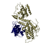
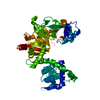
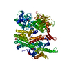
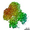
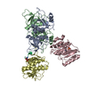
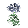
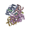
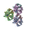
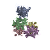
| Title | Structure of the cAMP responsive exchange factor Epac2 in its auto- inhibited state | ||||||
 Components Components | RAP GUANINE NUCLEOTIDE EXCHANGE FACTOR 4 | ||||||
 Keywords Keywords | REGULATION / EPAC2 / CAMP-GEF2 / CAMP / CYCLIC NUCLEOTIDE / GEF / EXCHANGE FACTOR / AUTO-INHIBITION / CDC25 HOMOLOGY DOMAIN | ||||||
| Function / homology |  Function and homology information Function and homology informationIntegrin signaling / Rap1 signalling / Regulation of insulin secretion / Glucagon-like Peptide-1 (GLP1) regulates insulin secretion / regulation of exocytosis / calcium-ion regulated exocytosis / hormone secretion / regulation of synaptic vesicle cycle / insulin secretion / small GTPase-mediated signal transduction ...Integrin signaling / Rap1 signalling / Regulation of insulin secretion / Glucagon-like Peptide-1 (GLP1) regulates insulin secretion / regulation of exocytosis / calcium-ion regulated exocytosis / hormone secretion / regulation of synaptic vesicle cycle / insulin secretion / small GTPase-mediated signal transduction / cAMP binding / guanyl-nucleotide exchange factor activity / hippocampal mossy fiber to CA3 synapse / positive regulation of insulin secretion / small GTPase binding / adenylate cyclase-modulating G protein-coupled receptor signaling pathway / adenylate cyclase-activating G protein-coupled receptor signaling pathway / protein-macromolecule adaptor activity / membrane / cytosol Similarity search - Function | ||||||
| Biological species |  | ||||||
| Method |  X-RAY DIFFRACTION / SYNCHROTRON / MOLECULAR REPLACEMENT / Resolution: 2.7 Å X-RAY DIFFRACTION / SYNCHROTRON / MOLECULAR REPLACEMENT / Resolution: 2.7 Å | ||||||
 Authors Authors | Rehmann, H. / Wittinghofer, A. / Bos, J.L. | ||||||
 Citation Citation | Journal: Nature / Year: 2006 Title: Structure of the Cyclic-AMP Responsive Exchange Factor Epac2 in its Auto-Inhibited State Authors: Rehmann, H. / Das, J. / Knipscheer, P. / Wittinghofer, A. / Bos, J.L. #1: Journal: Nat.Struct.Biol. / Year: 2002Title: Structure and Regulation of the Camp Binding Domains of Epac2 Authors: Rehmann, H. / Prakash, B. / Wolf, E. / Rueppel, A. / Derooij, J. / Bos, J.L. / Wittinghofer, A. | ||||||
| History |
|
- Structure visualization
| Structure viewer | Molecule: MolmilJmol/JSmol |
|---|
- Downloads & links
-Download
| PDBx/mmCIF format | 2byv.cif.gz | 193.9 KB | Display | PDBx/mmCIF format |
|---|---|---|---|---|
| PDB format | pdb2byv.ent.gz | 152 KB | Display | PDB format |
| PDBx/mmJSON format | 2byv.json.gz | Tree view | PDBx/mmJSON format | |
| Others |  Other downloads Other downloads |
-Validation report
| Arichive directory | https://data.pdbj.org/pub/pdb/validation_reports/by/2byvftp://data.pdbj.org/pub/pdb/validation_reports/by/2byv | HTTPS FTP |
|---|
-Related structure data

-Links
 PDBj
PDBj


- Assembly
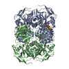
| Deposited unit | 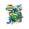
| ||||||||
|---|---|---|---|---|---|---|---|---|---|
| 1 |
| ||||||||
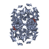
| Unit cell |
|
-Components
| #1: Protein | Mass: 114150.547 Da / Num. of mol.: 1 Source method: isolated from a genetically manipulated source Source: (gene. exp.)  |
|---|---|
| #2: Water | ChemComp-HOH /  Mass: 18.015 Da / Num. of mol.: 66 / Source method: isolated from a natural source / Formula: H2O Mass: 18.015 Da / Num. of mol.: 66 / Source method: isolated from a natural source / Formula: H2O |
-Experimental details
-Experiment
| Experiment | Method: X-RAY DIFFRACTION / Number of used crystals: 1 |
|---|
- Sample preparation
| Crystal | Density Matthews: 2.8 Å3/Da / Density % sol: 56 % |
|---|---|
| Crystal grow | pH: 7.5 Details: 100 MM BISTRISPROPANE7.5, 200 MM NANO3, 12% PEG 3350, pH 7.50 |
-Data collection
| Diffraction | Mean temperature: 100 K |
|---|---|
| Diffraction source | Source: SYNCHROTRON / Site: ESRF  / Beamline: ID14-4 / Wavelength: 0.976269 / Beamline: ID14-4 / Wavelength: 0.976269 |
| Detector | Detector: CCD / Date: Dec 14, 2004 |
| Radiation | Protocol: SINGLE WAVELENGTH / Monochromatic (M) / Laue (L): M / Scattering type: x-ray |
| Radiation wavelength | Wavelength: 0.976269 Å / Relative weight: 1 |
| Reflection | Resolution: 2.7→30 Å / Num. obs: 35428 / % possible obs: 98.6 % / Observed criterion σ(I): 0 / Redundancy: 7.3 % / Rmerge(I) obs: 0.06 / Net I/σ(I): 23.2 |
| Reflection shell | Resolution: 2.7→2.8 Å / Rmerge(I) obs: 0.3 / Mean I/σ(I) obs: 4.5 / % possible all: 96.9 |
- Processing
| Software |
| ||||||||||||||||||||||||||||||||||||||||||||||||||||||||||||||||||||||||||||||||||||||||||||||||||||||||||||||||||||||||||||||||||||||||||||||||||||||||||||||||||||||||||||||||||||||
|---|---|---|---|---|---|---|---|---|---|---|---|---|---|---|---|---|---|---|---|---|---|---|---|---|---|---|---|---|---|---|---|---|---|---|---|---|---|---|---|---|---|---|---|---|---|---|---|---|---|---|---|---|---|---|---|---|---|---|---|---|---|---|---|---|---|---|---|---|---|---|---|---|---|---|---|---|---|---|---|---|---|---|---|---|---|---|---|---|---|---|---|---|---|---|---|---|---|---|---|---|---|---|---|---|---|---|---|---|---|---|---|---|---|---|---|---|---|---|---|---|---|---|---|---|---|---|---|---|---|---|---|---|---|---|---|---|---|---|---|---|---|---|---|---|---|---|---|---|---|---|---|---|---|---|---|---|---|---|---|---|---|---|---|---|---|---|---|---|---|---|---|---|---|---|---|---|---|---|---|---|---|---|---|
| Refinement | Method to determine structure: MOLECULAR REPLACEMENT Starting model: PDB ENTRY 1O7F, PDB ENTRY 1BKD Resolution: 2.7→29.88 Å / Cor.coef. Fo:Fc: 0.921 / Cor.coef. Fo:Fc free: 0.881 / SU B: 14.86 / SU ML: 0.312 / Cross valid method: THROUGHOUT / ESU R: 0.885 / ESU R Free: 0.381 / Stereochemistry target values: MAXIMUM LIKELIHOOD Details: HYDROGENS HAVE BEEN ADDED IN THE RIDING POSITIONS. RESDIUES 1-14, 171-178, 465-476, 614-641, 725-731, 992-993 ARE DISORDERED
| ||||||||||||||||||||||||||||||||||||||||||||||||||||||||||||||||||||||||||||||||||||||||||||||||||||||||||||||||||||||||||||||||||||||||||||||||||||||||||||||||||||||||||||||||||||||
| Solvent computation | Ion probe radii: 0.8 Å / Shrinkage radii: 0.8 Å / VDW probe radii: 1.2 Å / Solvent model: MASK | ||||||||||||||||||||||||||||||||||||||||||||||||||||||||||||||||||||||||||||||||||||||||||||||||||||||||||||||||||||||||||||||||||||||||||||||||||||||||||||||||||||||||||||||||||||||
| Displacement parameters | Biso mean: 62.65 Å2
| ||||||||||||||||||||||||||||||||||||||||||||||||||||||||||||||||||||||||||||||||||||||||||||||||||||||||||||||||||||||||||||||||||||||||||||||||||||||||||||||||||||||||||||||||||||||
| Refinement step | Cycle: LAST / Resolution: 2.7→29.88 Å
| ||||||||||||||||||||||||||||||||||||||||||||||||||||||||||||||||||||||||||||||||||||||||||||||||||||||||||||||||||||||||||||||||||||||||||||||||||||||||||||||||||||||||||||||||||||||
| Refine LS restraints |
|