 Movie
Movie Controller

[English] 日本語
 Yorodumi
- PDB-2bpa: ATOMIC STRUCTURE OF SINGLE-STRANDED DNA BACTERIOPHAGE PHIX174 AND... -
+ Open data

- Basic information
| Entry | Database: PDB / ID: 2bpa | ||||||
|---|---|---|---|---|---|---|---|
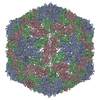
| Title | ATOMIC STRUCTURE OF SINGLE-STRANDED DNA BACTERIOPHAGE PHIX174 AND ITS FUNCTIONAL IMPLICATIONS | ||||||
 Components Components |
| ||||||
 Keywords Keywords | Virus/DNA /  PROTEIN-DNA COMPLEX / SINGLE STRAND / Icosahedral virus / Virus-DNA COMPLEX PROTEIN-DNA COMPLEX / SINGLE STRAND / Icosahedral virus / Virus-DNA COMPLEX | ||||||
| Function / homology |  Function and homology information Function and homology informationmodulation by virus of host process / T=1 icosahedral viral capsid / viral capsid / host cell cytoplasm / symbiont entry into host cell / virion attachment to host cell / structural molecule activity / DNA bindingSimilarity search - Function | ||||||
| Biological species |  Enterobacteria phage phiX174 (virus) Enterobacteria phage phiX174 (virus) | ||||||
| Method | X-RAY DIFFRACTION / Resolution: 3 Å | ||||||
 Authors Authors | McKenna, R. / Xia, D. / Willingmann, P. / Ilag, L.L. / Krishnaswamy, S. / Rossmann, M.G. / Olson, N.H. / Baker, T.S. / Incardona, N.L. | ||||||
 Citation Citation | Journal: Nature / Year: 1992 Title: Atomic structure of single-stranded DNA bacteriophage phi X174 and its functional implications. Authors: McKenna, R. / Xia, D. / Willingmann, P. / Ilag, L.L. / Krishnaswamy, S. / Rossmann, M.G. / Olson, N.H. / Baker, T.S. / Incardona, N.L. #1: Journal: Acta Crystallogr.,Sect.B / Year: 1992Title: Structure Determination of the Bacteriophage PhiX174 Authors: McKenna, R. / Xia, D. / Willingmann, P. / Ilag, L.L. / Rossmann, M.G. #2: Journal: J.Mol.Biol. / Year: 1994Title: Analysis of the Single-Stranded DNA Bacteriophage PhiX174 Refined at a Resolution of 3.0 A Authors: McKenna, R. / Ilag, L.L. / Rossmann, M.G. #3: Journal: J.Mol.Biol. / Year: 1990Title: Preliminary Investigation of the Phage PhiX174 Authors: Willingmann, P. / Krishnaswamy, S. / McKenna, R. / Smith, T.J. / Olson, N.H. / Rossmann, M.G. / Stow, P.L. / Incardona, N.L. #4: Journal: The Bacteriophages (The Viruses) / Year: 1988Title: Biology of the Bacteriophage PhiX174 Authors: Hayashi, M. / Adyama, A. / Delwood, L. / Richardson, D.L. / Hayashi, M.N. #5: Journal: Nature / Year: 1977Title: Nucleoside Sequence of Bacteriophage PhiX174 DNA Authors: Sanger, F. / Air, G.M. / Barrell, B.G. / Brown, N.L. / Coulson, A.R. / Fiddes, J.C. / Hutchinson, C.A. / Slocombe, P.M. / Smith, M. | ||||||
| History |
|
- Structure visualization
| Structure viewer | Molecule: MolmilJmol/JSmol |
|---|
- Downloads & links
-Download
| PDBx/mmCIF format | 2bpa.cif.gz | 144.6 KB | Display | PDBx/mmCIF format |
|---|---|---|---|---|
| PDB format | pdb2bpa.ent.gz | 111.2 KB | Display | PDB format |
| PDBx/mmJSON format | 2bpa.json.gz | Tree view | PDBx/mmJSON format | |
| Others |  Other downloads Other downloads |
-Validation report
| Arichive directory | https://data.pdbj.org/pub/pdb/validation_reports/bp/2bpaftp://data.pdbj.org/pub/pdb/validation_reports/bp/2bpa | HTTPS FTP |
|---|
-Related structure data
| Similar structure data |
|---|

-Links
 PDBj
PDBj


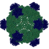
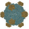
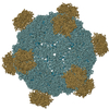
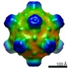
- Assembly
| Deposited unit | 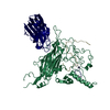
| ||||||||
|---|---|---|---|---|---|---|---|---|---|
| 1 | x 60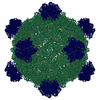
| ||||||||
| 2 |
| ||||||||
| 3 | x 5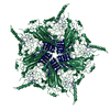
| ||||||||
| 4 | x 6
| ||||||||
| 5 | 
| ||||||||
| Unit cell |
| ||||||||
| Symmetry | Point symmetry: (Hermann–Mauguin notation: 532 / Schoenflies symbol: I (icosahedral)) |
-Components
| #1: DNA chain | Mass: 1497.052 Da / Num. of mol.: 1 / Source method: obtained synthetically |
|---|---|
| #2: Protein | Mass: 48423.375 Da / Num. of mol.: 1 Source method: isolated from a genetically manipulated source Source: (gene. exp.) Enterobacteria phage phiX174 (virus) / Genus: Microvirus / Species: Enterobacteria phage phiX174 sensu lato / Species (production host): Escherichia coli / Production host:  Escherichia coli C (bacteria) / Strain (production host): C / References: UniProt: P03641 Escherichia coli C (bacteria) / Strain (production host): C / References: UniProt: P03641 |
| #3: Protein | Mass: 19061.652 Da / Num. of mol.: 1 Source method: isolated from a genetically manipulated source Source: (gene. exp.) Enterobacteria phage phiX174 (virus) / Genus: Microvirus / Species: Enterobacteria phage phiX174 sensu lato / Species (production host): Escherichia coli / Production host: Escherichia coli C (bacteria) / Strain (production host): C / References: UniProt: P03643 |
| #4: Protein/peptide | Mass: 4107.821 Da / Num. of mol.: 1 Source method: isolated from a genetically manipulated source Source: (gene. exp.) Enterobacteria phage phiX174 (virus) / Genus: Microvirus / Species: Enterobacteria phage phiX174 sensu lato / Species (production host): Escherichia coli / Production host: Escherichia coli C (bacteria) / Strain (production host): C / References: UniProt: P69592 |
| #5: Water | ChemComp-HOH / Water Mass: 18.015 Da / Num. of mol.: 178 / Source method: isolated from a natural source / Formula: H2O Mass: 18.015 Da / Num. of mol.: 178 / Source method: isolated from a natural source / Formula: H2O |
| Sequence details | THE NUCLEIC ACID RESIDUES ARE LABELLED A AND C. WHILE THE PYRIMIDINE AND PURINE NATURES OF THE ...THE NUCLEIC ACID RESIDUES ARE LABELLED A AND C. WHILE THE PYRIMIDINE |
-Experimental details
-Experiment
| Experiment | Method: X-RAY DIFFRACTION |
|---|
- Sample preparation
| Crystal grow | *PLUS Method: vapor diffusion, hanging drop / Details: referred to J.Mol.Biol. 212.345-350 1990 | ||||||||||||||||||||||||||||||
|---|---|---|---|---|---|---|---|---|---|---|---|---|---|---|---|---|---|---|---|---|---|---|---|---|---|---|---|---|---|---|---|
| Components of the solutions | *PLUS
|
-Data collection
| Radiation | Protocol: SINGLE WAVELENGTH / Monochromatic (M) / Laue (L): M / Scattering type: x-ray |
|---|---|
| Radiation wavelength | Relative weight: 1 |
| Reflection | *PLUS Highest resolution: 2.7 Å / Lowest resolution: 9999 Å / Num. obs: 1000304 / Rmerge(I) obs: 0.129 |
| Reflection shell | *PLUS Highest resolution: 2.7 Å / Lowest resolution: 2.9 Å / % possible obs: 27 % / Num. unique obs: 92108 |
- Processing
| Software | Name: PROLSQ / Classification: refinement | |||||||||||||||||||||||||||||||||||||||||||||||||||||||||||||||
|---|---|---|---|---|---|---|---|---|---|---|---|---|---|---|---|---|---|---|---|---|---|---|---|---|---|---|---|---|---|---|---|---|---|---|---|---|---|---|---|---|---|---|---|---|---|---|---|---|---|---|---|---|---|---|---|---|---|---|---|---|---|---|---|---|
| Refinement | Resolution: 3→6 Å / Rfactor obs: 0.209 / σ(F): 5 Details: ALL 175 AMINO ACIDS OF GPG, ALL 426 AMINO ACIDS OF GPF AND ALL BUT THE FIRST AMINO ACID OF GPJ WERE BUILT. ALSO FOUR NUCLEOTIDES (A 0-4) WERE BUILT, ALTHOUGH THE CHOICE OF NUCLEOTIDE BASES WERE ARBITRARY. | |||||||||||||||||||||||||||||||||||||||||||||||||||||||||||||||
| Refinement step | Cycle: LAST / Resolution: 3→6 Å
| |||||||||||||||||||||||||||||||||||||||||||||||||||||||||||||||
| Refine LS restraints |
| |||||||||||||||||||||||||||||||||||||||||||||||||||||||||||||||
| Refinement | *PLUS Highest resolution: 3 Å / Lowest resolution: 6 Å / σ(F): 5 / Rfactor obs: 0.209 | |||||||||||||||||||||||||||||||||||||||||||||||||||||||||||||||
| Solvent computation | *PLUS | |||||||||||||||||||||||||||||||||||||||||||||||||||||||||||||||
| Displacement parameters | *PLUS | |||||||||||||||||||||||||||||||||||||||||||||||||||||||||||||||
| Refine LS restraints | *PLUS
|
