 Movie
Movie Controller


+ Open data

- Basic information
| Entry | Database: PDB / ID: 1a6c | |||||||||
|---|---|---|---|---|---|---|---|---|---|---|
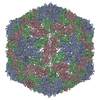
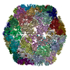
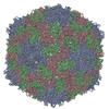
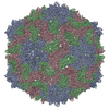
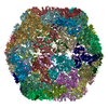
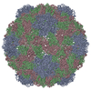
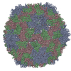
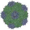
| Title | STRUCTURE OF TOBACCO RINGSPOT VIRUS | |||||||||
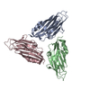
 Components Components | TOBACCO RINGSPOT VIRUS CAPSID PROTEIN | |||||||||
 Keywords Keywords | VIRUS / TRSV / NEPOVIRUS / VIRUS STRUCTURE / VIRUS EVOLUTION / PICORNAVIRUS SUPERFAMILY / VIRUS CAPSID PROTEIN / Icosahedral virus | |||||||||
| Function / homology |  Function and homology information Function and homology information | |||||||||
| Biological species |  Tobacco ringspot virus Tobacco ringspot virus | |||||||||
| Method |  X-RAY DIFFRACTION / SYNCHROTRON / MOLECULAR REPLACEMENT / Resolution: 3.5 Å X-RAY DIFFRACTION / SYNCHROTRON / MOLECULAR REPLACEMENT / Resolution: 3.5 Å | |||||||||
 Authors Authors | Johnson, J.E. / Chandrasekar, V. | |||||||||
 Citation Citation | Journal: Structure / Year: 1998 Title: The structure of tobacco ringspot virus: a link in the evolution of icosahedral capsids in the picornavirus superfamily. Authors: Chandrasekar, V. / Johnson, J.E. #1: Journal: Virology / Year: 1995Title: Expression of Tobacco Ringspot Virus Capsid Protein and Satellite RNA in Insect Cells and Three-Dimensional Structure of Tobacco Ringspot Virus-Like Particles Authors: Singh, S. / Rothnagel, R. / Prasad, B.V. / Buckley, B. #2: Journal: Virus Res. / Year: 1995Title: Erratum. Nucleotide Sequence and in Vitro Expression of the Capsid Protein Gene of Tobacco Ringspot Virus Authors: Buckley, B. / Silva, S. / Singh, S. #3: Journal: Virus Res. / Year: 1993Title: Nucleotide Sequence and in Vitro Expression of the Capsid Protein Gene of Tobacco Ringspot Virus Authors: Buckley, B. / Silva, S. / Singh, S. #4: Journal: Science / Year: 1989Title: Protein-RNA Interactions in an Icosahedral Virus at 3.0 A Resolution Authors: Chen, Z.G. / Stauffacher, C. / Li, Y. / Schmidt, T. / Bomu, W. / Kamer, G. / Shanks, M. / Lomonossoff, G. / Johnson, J.E. #5: Journal: Nature / Year: 1985Title: Structure of a Human Common Cold Virus and Functional Relationship to Other Picornaviruses Authors: Rossmann, M.G. / Arnold, E. / Erickson, J.W. / Frankenberger, E.A. / Griffith, J.P. / Hecht, H.J. / Johnson, J.E. / Kamer, G. / Luo, M. / Mosser, A.G. / Rueckert, R.R. / Sherry, B. / Vriend, G. | |||||||||
| History |
|
- Structure visualization
| Structure viewer | Molecule: MolmilJmol/JSmol |
|---|
- Downloads & links
-Download
| PDBx/mmCIF format | 1a6c.cif.gz | 101.8 KB | Display | PDBx/mmCIF format |
|---|---|---|---|---|
| PDB format | pdb1a6c.ent.gz | 76.4 KB | Display | PDB format |
| PDBx/mmJSON format | 1a6c.json.gz | Tree view | PDBx/mmJSON format | |
| Others |  Other downloads Other downloads |
-Validation report
| Arichive directory | https://data.pdbj.org/pub/pdb/validation_reports/a6/1a6cftp://data.pdbj.org/pub/pdb/validation_reports/a6/1a6c | HTTPS FTP |
|---|
-Related structure data
| Similar structure data |
|---|

-Links
 PDBj
PDBj- Assembly
| Deposited unit | 
| |||||||||||||||||||||||||||||||||||||||||||||||||||||||||||||||||||||||||||||||||||||||||||||
|---|---|---|---|---|---|---|---|---|---|---|---|---|---|---|---|---|---|---|---|---|---|---|---|---|---|---|---|---|---|---|---|---|---|---|---|---|---|---|---|---|---|---|---|---|---|---|---|---|---|---|---|---|---|---|---|---|---|---|---|---|---|---|---|---|---|---|---|---|---|---|---|---|---|---|---|---|---|---|---|---|---|---|---|---|---|---|---|---|---|---|---|---|---|---|
| 1 | x 60
| |||||||||||||||||||||||||||||||||||||||||||||||||||||||||||||||||||||||||||||||||||||||||||||
| 2 |
| |||||||||||||||||||||||||||||||||||||||||||||||||||||||||||||||||||||||||||||||||||||||||||||
| 3 | x 5
| |||||||||||||||||||||||||||||||||||||||||||||||||||||||||||||||||||||||||||||||||||||||||||||
| 4 | x 6
| |||||||||||||||||||||||||||||||||||||||||||||||||||||||||||||||||||||||||||||||||||||||||||||
| 5 | 
| |||||||||||||||||||||||||||||||||||||||||||||||||||||||||||||||||||||||||||||||||||||||||||||
| 6 | x 30
| |||||||||||||||||||||||||||||||||||||||||||||||||||||||||||||||||||||||||||||||||||||||||||||
| Unit cell |
| |||||||||||||||||||||||||||||||||||||||||||||||||||||||||||||||||||||||||||||||||||||||||||||
| Symmetry | Point symmetry: (Hermann–Mauguin notation: 532 / Schoenflies symbol: I (icosahedral)) | |||||||||||||||||||||||||||||||||||||||||||||||||||||||||||||||||||||||||||||||||||||||||||||
| Noncrystallographic symmetry (NCS) | NCS oper:
|
-Components
| #1: Protein | Mass: 56993.648 Da / Num. of mol.: 1 / Source method: isolated from a natural source / Source: (natural) Tobacco ringspot virus / Genus: Nepovirus / Strain: XANTHI-NC / References: UniProt: Q88894 |
|---|
-Experimental details
-Experiment
| Experiment | Method: X-RAY DIFFRACTION / Number of used crystals: 27 |
|---|
- Sample preparation
| Crystal | Density Matthews: 3.2 Å3/Da |
|---|---|
| Crystal grow | Method: vapor diffusion, hanging drop / pH: 6 Details: VIRUS WAS CRYSTALLIZED USING HANGING DROP SETTING FROM RESERVOIR BUFFER CONTAINING 2-3% (W/V) PEG 3350, 1MM SODIUM AZIDE AND 0.125 M POTASSIUM PHOSPHATE, PH 6.5, pH 6.0, vapor diffusion - hanging drop |
| Crystal grow | *PLUS Method: unknown |
| Components of the solutions | *PLUS Conc.: 2-3 % / Common name: PEG MW 3350 |
-Data collection
| Diffraction | Mean temperature: 300 K |
|---|---|
| Diffraction source | Source: SYNCHROTRON / Site: CHESS  / Beamline: F1 / Wavelength: 0.918 / Beamline: F1 / Wavelength: 0.918 |
| Detector | Type: FUJI / Detector: IMAGE PLATE / Date: Apr 1, 1994 / Details: MIRRORS |
| Radiation | Monochromator: SI(111) / Monochromatic (M) / Laue (L): M / Scattering type: x-ray |
| Radiation wavelength | Wavelength: 0.918 Å / Relative weight: 1 |
| Reflection | Resolution: 3.5→30 Å / Num. obs: 92395 / % possible obs: 18 % / Observed criterion σ(I): 4 / Redundancy: 1.2 % / Rmerge(I) obs: 0.084 / Rsym value: 0.15 |
| Reflection shell | Resolution: 3.5→4 Å / % possible all: 10 |
- Processing
| Software |
| ||||||||||||||||||||||||||||||||||||||||||||||||||||||||||||
|---|---|---|---|---|---|---|---|---|---|---|---|---|---|---|---|---|---|---|---|---|---|---|---|---|---|---|---|---|---|---|---|---|---|---|---|---|---|---|---|---|---|---|---|---|---|---|---|---|---|---|---|---|---|---|---|---|---|---|---|---|---|
| Refinement | Method to determine structure: MOLECULAR REPLACEMENT Starting model: STRUCTURE OF COMOVIRUS CPMV Resolution: 3.5→8 Å / Data cutoff high absF: 100000 / Data cutoff low absF: 0.1 / σ(F): 1 Details: THE STRUCTURE WAS REFINED TO 3.5 ANGSTROMS RESOLUTION USING ONLY 27% OF THE THEORETICALLY POSSIBLE UNIQUE REFLECTIONS BETWEEN 8.0 AND 3.5 ANGSTROMS. THERE ARE STILL SOME BOND ANGLES AND BOND ...Details: THE STRUCTURE WAS REFINED TO 3.5 ANGSTROMS RESOLUTION USING ONLY 27% OF THE THEORETICALLY POSSIBLE UNIQUE REFLECTIONS BETWEEN 8.0 AND 3.5 ANGSTROMS. THERE ARE STILL SOME BOND ANGLES AND BOND LENGTHS IN THE CRYSTAL STRUCTURE WHICH VARY BY GREATER THAN 3.0 RMSD FROM THEIR CORRESPONDING E & H VALUES. THESE HAVE NOT BEEN FIXED AT PRESENT.
| ||||||||||||||||||||||||||||||||||||||||||||||||||||||||||||
| Displacement parameters | Biso mean: 15 Å2 | ||||||||||||||||||||||||||||||||||||||||||||||||||||||||||||
| Refinement step | Cycle: LAST / Resolution: 3.5→8 Å
| ||||||||||||||||||||||||||||||||||||||||||||||||||||||||||||
| Refine LS restraints |
| ||||||||||||||||||||||||||||||||||||||||||||||||||||||||||||
| LS refinement shell | Resolution: 3.5→3.64 Å / Total num. of bins used: 8 /
| ||||||||||||||||||||||||||||||||||||||||||||||||||||||||||||
| Xplor file |
|
