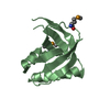
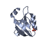
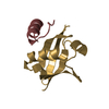
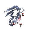
Entry Database : PDB / ID : 2awuTitle Synapse associated protein 97 PDZ2 domain variant C378G AHH Synapse-associated protein 97 Keywords / / Function / homology Function Domain/homology Component
/ / / / / / / / / / / / / / / / / / / / / / / / / / / / / / / / / / / / / / / / / / / / / / / / / / / / / / / / / / / / / / / / / / / / / / / / / / / / / / / / / / / / / / / / / / / / / / / / / / / / / / / / / / / / / / / / / / / / / / / / / / / / / / / / / / / / / / / / / / / / / / / / / / / / / / / Biological species Rattus norvegicus (Norway rat)Method / / / Resolution : 2.44 Å Authors Von Ossowski, I. / Oksanen, E. / Von Ossowski, L. / Cai, C. / Sundberg, M. / Goldman, A. / Keinanen, K. Journal : Febs J. / Year : 2006Title : Crystal structure of the second PDZ domain of SAP97 in complex with a GluR-A C-terminal peptideAuthors : von Ossowski, I. / Oksanen, E. / von Ossowski, L. / Cai, C. / Sundberg, M. / Goldman, A. / Keinanen, K. History Deposition Sep 2, 2005 Deposition site / Processing site Revision 1.0 Aug 29, 2006 Provider / Type Revision 1.1 May 1, 2008 Group Revision 1.2 Jul 13, 2011 Group Revision 1.3 Oct 11, 2017 Group / Category / Item / _software.nameRevision 1.4 Nov 10, 2021 Group / Database references / Category / diffrn_source / struct_ref_seq_difItem _database_2.pdbx_DOI / _database_2.pdbx_database_accession ... _database_2.pdbx_DOI / _database_2.pdbx_database_accession / _diffrn_source.pdbx_synchrotron_site / _struct_ref_seq_dif.details Revision 1.5 May 29, 2024 Group / Category / chem_comp_bond
Show all Show less
 Movie
Movie Controller


 Open data
 Basic information
 Components
 Keywords
 Function and homology information
 X-RAY DIFFRACTION /
  Authors
 Citation
 Structure visualization
 Downloads & links
 Other downloads
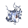


 PDBj
PDBj


 Assembly


 Mass: 18.015 Da / Num. of mol.: 74 / Source method: isolated from a natural source / Formula: H2O
 Sample preparation
 / Beamline: BW7A / Wavelength: 1.519 Å
 Processing