 Movie
Movie Controller


+ Open data

- Basic information
| Entry | Database: PDB / ID: 1z3f | ||||||||||||||||||
|---|---|---|---|---|---|---|---|---|---|---|---|---|---|---|---|---|---|---|---|
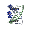
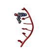
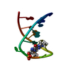
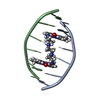
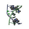
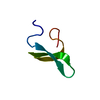
| Title | Structure of ellipticine in complex with a 6-bp DNA | ||||||||||||||||||
 Components Components | 5'-D(* Keywords KeywordsDNA / ANTICANCER / DNA BINDING / DRUG DESIGN / ELLIPTICINE / INTERCALATOR | Function / homology | : / ELLIPTICINE / DNA |  Function and homology information Function and homology informationMethod |  X-RAY DIFFRACTION / SYNCHROTRON / MOLECULAR REPLACEMENT / Resolution: 1.5 Å X-RAY DIFFRACTION / SYNCHROTRON / MOLECULAR REPLACEMENT / Resolution: 1.5 Å  Authors AuthorsCanals, A. / Purciolas, M. / Aymami, J. / Coll, M. |  CitationJournal: Acta Crystallogr.,Sect.D / Year: 2005 CitationJournal: Acta Crystallogr.,Sect.D / Year: 2005Title: The anticancer agent ellipticine unwinds DNA by intercalative binding in an orientation parallel to base pairs. Authors: Canals, A. / Purciolas, M. / Aymami, J. / Coll, M. History |
|
- Structure visualization
| Structure viewer | Molecule: MolmilJmol/JSmol |
|---|
- Downloads & links
-Download
| PDBx/mmCIF format | 1z3f.cif.gz | 24.9 KB | Display | PDBx/mmCIF format |
|---|---|---|---|---|
| PDB format | pdb1z3f.ent.gz | 17.4 KB | Display | PDB format |
| PDBx/mmJSON format | 1z3f.json.gz | Tree view | PDBx/mmJSON format | |
| Others |  Other downloads Other downloads |
-Validation report
| Arichive directory | https://data.pdbj.org/pub/pdb/validation_reports/z3/1z3fftp://data.pdbj.org/pub/pdb/validation_reports/z3/1z3f | HTTPS FTP |
|---|
-Related structure data
| Similar structure data |
|---|


-Links
 PDBj
PDBj


- Assembly
| Deposited unit | 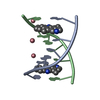
| ||||||||
|---|---|---|---|---|---|---|---|---|---|
| 1 |
| ||||||||
| Unit cell |
|
-Components
| #1: DNA chain | Mass: 1809.217 Da / Num. of mol.: 2 / Source method: obtained synthetically #2: Chemical |   Mass: 247.314 Da / Num. of mol.: 2 / Source method: obtained synthetically / Formula: C17H15N2 / Comment: alkaloid*YM Mass: 247.314 Da / Num. of mol.: 2 / Source method: obtained synthetically / Formula: C17H15N2 / Comment: alkaloid*YM#3: Chemical |   Mass: 58.933 Da / Num. of mol.: 3 / Source method: obtained synthetically / Formula: Co Mass: 58.933 Da / Num. of mol.: 3 / Source method: obtained synthetically / Formula: Co#4: Water | ChemComp-HOH / |  Mass: 18.015 Da / Num. of mol.: 27 / Source method: isolated from a natural source / Formula: H2O Mass: 18.015 Da / Num. of mol.: 27 / Source method: isolated from a natural source / Formula: H2O |
|---|
-Experimental details
-Experiment
| Experiment | Method: X-RAY DIFFRACTION / Number of used crystals: 1 |
|---|
- Sample preparation
| Crystal | Density Matthews: 1.62 Å3/Da / Density % sol: 23.27 % | ||||||||||||||||||||||||||||||||||||||||||||||||
|---|---|---|---|---|---|---|---|---|---|---|---|---|---|---|---|---|---|---|---|---|---|---|---|---|---|---|---|---|---|---|---|---|---|---|---|---|---|---|---|---|---|---|---|---|---|---|---|---|---|
| Crystal grow | Temperature: 293 K / Method: vapor diffusion, hanging drop / pH: 5.5 Details: MPD, sodium cacodylate, cobalt hexammine, sodium chloride, potassium chloride, pH 5.5, VAPOR DIFFUSION, HANGING DROP, temperature 293K | ||||||||||||||||||||||||||||||||||||||||||||||||
| Components of the solutions |
|
-Data collection
| Diffraction | Mean temperature: 120 K |
|---|---|
| Diffraction source | Source: SYNCHROTRON / Site: ESRF  / Beamline: ID13 / Wavelength: 0.976 Å / Beamline: ID13 / Wavelength: 0.976 Å |
| Detector | Type: MARRESEARCH / Detector: CCD / Date: Jun 14, 2004 / Details: microfocus beamline |
| Radiation | Monochromator: liq. N2 cooled Si-111 double monochromator / Protocol: SINGLE WAVELENGTH / Monochromatic (M) / Laue (L): M / Scattering type: x-ray |
| Radiation wavelength | Wavelength: 0.976 Å / Relative weight: 1 |
| Reflection | Resolution: 1.5→16.66 Å / Num. all: 4436 / Num. obs: 4420 / % possible obs: 99.6 % / Observed criterion σ(F): 1 / Observed criterion σ(I): 1 |
| Reflection shell | Resolution: 1.5→1.54 Å / % possible all: 99.7 |
- Processing
| Software |
| |||||||||||||||||||||||||||||||||||||||||||||||||||||||||||||||||||||||||||
|---|---|---|---|---|---|---|---|---|---|---|---|---|---|---|---|---|---|---|---|---|---|---|---|---|---|---|---|---|---|---|---|---|---|---|---|---|---|---|---|---|---|---|---|---|---|---|---|---|---|---|---|---|---|---|---|---|---|---|---|---|---|---|---|---|---|---|---|---|---|---|---|---|---|---|---|---|
| Refinement | Method to determine structure: MOLECULAR REPLACEMENT / Resolution: 1.5→16.66 Å / Cor.coef. Fo:Fc: 0.969 / Cor.coef. Fo:Fc free: 0.971 / SU B: 6.257 / SU ML: 0.104 / Cross valid method: THROUGHOUT / σ(F): 0 / ESU R: 0.156 / ESU R Free: 0.1 / Stereochemistry target values: MAXIMUM LIKELIHOOD / Details: HYDROGENS HAVE BEEN ADDED IN THE RIDING POSITIONS
| |||||||||||||||||||||||||||||||||||||||||||||||||||||||||||||||||||||||||||
| Solvent computation | Ion probe radii: 0.8 Å / Shrinkage radii: 0.8 Å / VDW probe radii: 1.2 Å / Solvent model: MASK | |||||||||||||||||||||||||||||||||||||||||||||||||||||||||||||||||||||||||||
| Displacement parameters | Biso mean: 30.952 Å2
| |||||||||||||||||||||||||||||||||||||||||||||||||||||||||||||||||||||||||||
| Refinement step | Cycle: LAST / Resolution: 1.5→16.66 Å
| |||||||||||||||||||||||||||||||||||||||||||||||||||||||||||||||||||||||||||
| Refine LS restraints |
| |||||||||||||||||||||||||||||||||||||||||||||||||||||||||||||||||||||||||||
| LS refinement shell | Resolution: 1.5→1.539 Å / Total num. of bins used: 20
|
