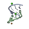
 Movie
Movie Controller

[English] 日本語
 Yorodumi
- PDB-2gjb: Crosslinking of DNA duplexes: X-ray crystal structure of an unsub... -
+ Open data

- Basic information
| Entry | Database: PDB / ID: 2gjb | ||||||||||||||||||
|---|---|---|---|---|---|---|---|---|---|---|---|---|---|---|---|---|---|---|---|
| Title | Crosslinking of DNA duplexes: X-ray crystal structure of an unsubstituted bisacridine with the oligonucleotide d(CGTACG) | ||||||||||||||||||
 Components Components | 5'-D(* Keywords KeywordsDNA / DNA duplex / bis-intercalator | Function / homology | Chem-BMO / : / DNA |  Function and homology information Function and homology informationMethod |  X-RAY DIFFRACTION / SYNCHROTRON / MOLECULAR REPLACEMENT / Resolution: 2.2 Å X-RAY DIFFRACTION / SYNCHROTRON / MOLECULAR REPLACEMENT / Resolution: 2.2 Å  Authors AuthorsGan, Y. / Cardin, C.J. / Denny, W.A. |  CitationJournal: To be Published CitationJournal: To be PublishedTitle: Crosslinking of DNA duplexes: X-ray crystal structure of an unsubstituted bisacridine with oligonucleotide d(CGTACG) Authors: Gan, Y. / Cardin, C.J. / Denny, W.A. History |
|
- Structure visualization
| Structure viewer | Molecule: MolmilJmol/JSmol |
|---|
- Downloads & links
-Download
| PDBx/mmCIF format | 2gjb.cif.gz | 14.6 KB | Display | PDBx/mmCIF format |
|---|---|---|---|---|
| PDB format | pdb2gjb.ent.gz | 8.1 KB | Display | PDB format |
| PDBx/mmJSON format | 2gjb.json.gz | Tree view | PDBx/mmJSON format | |
| Others |  Other downloads Other downloads |
-Validation report
| Arichive directory | https://data.pdbj.org/pub/pdb/validation_reports/gj/2gjbftp://data.pdbj.org/pub/pdb/validation_reports/gj/2gjb | HTTPS FTP |
|---|
-Related structure data
| Related structure data | |
|---|---|
| Similar structure data |
-Links
 PDBj
PDBj

- Assembly
| Deposited unit | 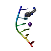
| ||||||||
|---|---|---|---|---|---|---|---|---|---|
| 1 | 
| ||||||||
| Unit cell |
| ||||||||
| Components on special symmetry positions |
| ||||||||
| Details | The second part of the biological assembly is generated by the two fold axis |
-Components
| #1: DNA chain | Mass: 1809.217 Da / Num. of mol.: 1 / Source method: obtained synthetically |
|---|---|
| #2: Chemical | ChemComp-K /   Mass: 39.098 Da / Num. of mol.: 1 / Source method: obtained synthetically / Formula: K Mass: 39.098 Da / Num. of mol.: 1 / Source method: obtained synthetically / Formula: K |
| #3: Chemical | ChemComp-BMO / 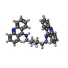  Mass: 485.622 Da / Num. of mol.: 1 / Source method: obtained synthetically / Formula: C32H31N5 Mass: 485.622 Da / Num. of mol.: 1 / Source method: obtained synthetically / Formula: C32H31N5 |
| #4: Water | ChemComp-HOH /  Mass: 18.015 Da / Num. of mol.: 1 / Source method: isolated from a natural source / Formula: H2O Mass: 18.015 Da / Num. of mol.: 1 / Source method: isolated from a natural source / Formula: H2O |
-Experimental details
-Experiment
| Experiment | Method: X-RAY DIFFRACTION / Number of used crystals: 1 |
|---|
- Sample preparation
| Crystal | Density Matthews: 2 Å3/Da / Density % sol: 38.53 % | ||||||||||||||||||||||||||||||||||||
|---|---|---|---|---|---|---|---|---|---|---|---|---|---|---|---|---|---|---|---|---|---|---|---|---|---|---|---|---|---|---|---|---|---|---|---|---|---|
| Crystal grow | Temperature: 293 K / Method: vapor diffusion, sitting drop / pH: 7 Details: The crystals grow in ten days from sitting drops containing 2mM DNA, 12mM spermine, 80mM strontium chloride, 20mM magnesium chloride, 10%MPD, pH 7.0, VAPOR DIFFUSION, SITTING DROP, temperature 293K | ||||||||||||||||||||||||||||||||||||
| Components of the solutions |
|
-Data collection
| Diffraction | Mean temperature: 100 K |
|---|---|
| Diffraction source | Source: SYNCHROTRON / Site: EMBL/DESY, HAMBURG  / Beamline: X12 / Wavelength: 1 Å / Beamline: X12 / Wavelength: 1 Å |
| Radiation | Protocol: SINGLE WAVELENGTH / Monochromatic (M) / Laue (L): M / Scattering type: x-ray |
| Radiation wavelength | Wavelength: 1 Å / Relative weight: 1 |
| Reflection | Resolution: 1.9→22.3 Å / Num. obs: 1472 / % possible obs: 99.3 % / Observed criterion σ(F): 2 / Observed criterion σ(I): 2 / Redundancy: 20.1 % / Biso Wilson estimate: 57.431 Å2 / Rmerge(I) obs: 0.038 / Net I/σ(I): 49.4 |
| Reflection shell | Resolution: 1.9→2 Å / Redundancy: 15.3 % / Rmerge(I) obs: 0.312 / Mean I/σ(I) obs: 9 / Num. unique all: 204 / % possible all: 100 |
- Processing
| Software |
| |||||||||||||||||||||||||||||||||||||||||||||||||||||||
|---|---|---|---|---|---|---|---|---|---|---|---|---|---|---|---|---|---|---|---|---|---|---|---|---|---|---|---|---|---|---|---|---|---|---|---|---|---|---|---|---|---|---|---|---|---|---|---|---|---|---|---|---|---|---|---|---|
| Refinement | Method to determine structure: MOLECULAR REPLACEMENT Starting model: NDB ENTRY DD0018 Resolution: 2.2→8 Å / Cor.coef. Fo:Fc: 0.943 / Cor.coef. Fo:Fc free: 0.96 / SU B: 5.648 / SU ML: 0.15 / Cross valid method: THROUGHOUT / ESU R: 0.398 / ESU R Free: 0.272 / Stereochemistry target values: MAXIMUM LIKELIHOOD / Details: HYDROGENS HAVE BEEN ADDED IN THE RIDING POSITIONS
| |||||||||||||||||||||||||||||||||||||||||||||||||||||||
| Solvent computation | Ion probe radii: 0.8 Å / Shrinkage radii: 0.8 Å / VDW probe radii: 1.2 Å / Solvent model: MASK | |||||||||||||||||||||||||||||||||||||||||||||||||||||||
| Displacement parameters | Biso mean: 37.613 Å2
| |||||||||||||||||||||||||||||||||||||||||||||||||||||||
| Refinement step | Cycle: LAST / Resolution: 2.2→8 Å
| |||||||||||||||||||||||||||||||||||||||||||||||||||||||
| Refine LS restraints |
| |||||||||||||||||||||||||||||||||||||||||||||||||||||||
| LS refinement shell | Resolution: 2.2→2.253 Å / Total num. of bins used: 20
|
