 Movie
Movie Controller

[English] 日本語
 Yorodumi
- PDB-4hig: Ultrahigh-resolution crystal structure of Z-DNA in complex with M... -
+ Open data

- Basic information
| Entry | Database: PDB / ID: 4hig | ||||||||||||||||||||
|---|---|---|---|---|---|---|---|---|---|---|---|---|---|---|---|---|---|---|---|---|---|
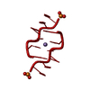
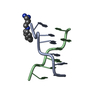
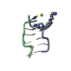
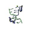
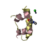
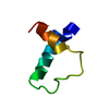
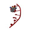
| Title | Ultrahigh-resolution crystal structure of Z-DNA in complex with Mn2+ ion. | ||||||||||||||||||||
 Components Components | DNA (5'-D(* Keywords KeywordsDNA / self-complementary DNA / Z-DNA | Function / homology | : / SPERMINE (FULLY PROTONATED FORM) / DNA |  Function and homology information Function and homology informationBiological species | synthetic construct (others) | Method |  X-RAY DIFFRACTION / SYNCHROTRON / MOLECULAR REPLACEMENT / Resolution: 0.75 Å X-RAY DIFFRACTION / SYNCHROTRON / MOLECULAR REPLACEMENT / Resolution: 0.75 Å  Authors AuthorsDrozdzal, P. / Gilski, M. / Kierzek, R. / Lomozik, L. / Jaskolski, M. |  CitationJournal: Acta Crystallogr.,Sect.D / Year: 2013 CitationJournal: Acta Crystallogr.,Sect.D / Year: 2013Title: Ultrahigh-resolution crystal structures of Z-DNA in complex with Mn(2+) and Zn(2+) ions. Authors: Drozdzal, P. / Gilski, M. / Kierzek, R. / Lomozik, L. / Jaskolski, M. #1: Journal: Nucleic Acids Res. / Year: 2011Title: High regularity of Z-DNA revealed by ultra high-resolution crystal structure at 0.55 A. Authors: Brzezinski, K. / Brzuszkiewicz, A. / Dauter, M. / Kubicki, M. / Jaskolski, M. / Dauter, Z. #2: Journal: Acta Crystallogr.,Sect.D / Year: 2001Title: Anomalous signal of phosphorus used for phasing DNA oligomer: importance of data redundancy. Authors: Dauter, Z. / Adamiak, D.A. History |
|
- Structure visualization
| Structure viewer | Molecule: MolmilJmol/JSmol |
|---|
- Downloads & links
-Download
| PDBx/mmCIF format | 4hig.cif.gz | 28.3 KB | Display | PDBx/mmCIF format |
|---|---|---|---|---|
| PDB format | pdb4hig.ent.gz | 18.9 KB | Display | PDB format |
| PDBx/mmJSON format | 4hig.json.gz | Tree view | PDBx/mmJSON format | |
| Others |  Other downloads Other downloads |
-Validation report
| Arichive directory | https://data.pdbj.org/pub/pdb/validation_reports/hi/4higftp://data.pdbj.org/pub/pdb/validation_reports/hi/4hig | HTTPS FTP |
|---|
-Related structure data
| Related structure data | 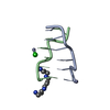 4hifC  1i0tS C: citing same article ( S: Starting model for refinement |
|---|---|
| Similar structure data |


-Links
 PDBj
PDBj

- Assembly
| Deposited unit | 
| ||||||||
|---|---|---|---|---|---|---|---|---|---|
| 1 |
| ||||||||
| Unit cell |
|
-Components
| #1: DNA chain | Mass: 1810.205 Da / Num. of mol.: 2 / Source method: obtained synthetically / Details: Synthetic construct / Source: (synth.) synthetic construct (others) #2: Chemical | ChemComp-SPK / |   Mass: 206.372 Da / Num. of mol.: 1 / Source method: obtained synthetically / Formula: C10H30N4 Mass: 206.372 Da / Num. of mol.: 1 / Source method: obtained synthetically / Formula: C10H30N4#3: Chemical | ChemComp-MN / |   Mass: 54.938 Da / Num. of mol.: 1 / Source method: obtained synthetically / Formula: Mn Mass: 54.938 Da / Num. of mol.: 1 / Source method: obtained synthetically / Formula: Mn#4: Water | ChemComp-HOH / |  Mass: 18.015 Da / Num. of mol.: 92 / Source method: isolated from a natural source / Formula: H2O Mass: 18.015 Da / Num. of mol.: 92 / Source method: isolated from a natural source / Formula: H2O |
|---|
-Experimental details
-Experiment
| Experiment | Method: X-RAY DIFFRACTION / Number of used crystals: 2 |
|---|
- Sample preparation
| Crystal | Density Matthews: 1.59 Å3/Da / Density % sol: 22.7 % |
|---|---|
| Crystal grow | Temperature: 292 K / pH: 6 Details: 1.5 MM DNA WATER SOLUTION MIXED 1:1 V/V WITH 10% MPD, 12 MM SPERMINE TETRA-HCL, 12 MM NACL, 80 MM KCL AND EQUILIBRATED AGAINST 35% MPD, PH 6. VAPOR DIFFUSION, HANGING DROP METHOD, ...Details: 1.5 MM DNA WATER SOLUTION MIXED 1:1 V/V WITH 10% MPD, 12 MM SPERMINE TETRA-HCL, 12 MM NACL, 80 MM KCL AND EQUILIBRATED AGAINST 35% MPD, PH 6. VAPOR DIFFUSION, HANGING DROP METHOD, TEMPERATURE 292K. FOR MN2+ SOAKING, A CRYSTAL WAS PLACED IN 0.002 ML OF THE RESERVOIR SOLUTION MIXED WITH 0.002 ML OF 5 MM MNCL2 FOR ONE WEEK. |
-Data collection
| Diffraction |
| ||||||||||||||||||
|---|---|---|---|---|---|---|---|---|---|---|---|---|---|---|---|---|---|---|---|
| Diffraction source |
| ||||||||||||||||||
| Detector |
| ||||||||||||||||||
| Radiation |
| ||||||||||||||||||
| Radiation wavelength |
| ||||||||||||||||||
| Reflection | Resolution: 0.75→25.57 Å / Num. obs: 47730 / % possible obs: 79.9 % / Observed criterion σ(I): -3 / Redundancy: 3.28 % / Biso Wilson estimate: 4.756 Å2 / Rmerge(I) obs: 0.053 / Net I/σ(I): 19.28 | ||||||||||||||||||
| Reflection shell | Resolution: 0.75→0.77 Å / Redundancy: 1.15 % / Rmerge(I) obs: 0.038 / Mean I/σ(I) obs: 11.39 / % possible all: 10.6 |

- Processing
| Software |
| |||||||||||||||||||||||||||||||||
|---|---|---|---|---|---|---|---|---|---|---|---|---|---|---|---|---|---|---|---|---|---|---|---|---|---|---|---|---|---|---|---|---|---|---|
| Refinement | Method to determine structure: MOLECULAR REPLACEMENT Starting model: 1I0T Resolution: 0.75→25.57 Å / Num. parameters: 3409 / Num. restraintsaints: 1139 / Cross valid method: FREE R Details: THE REFINEMENT WAS CARRIED OUT AGAINST SEPARATE BIJVOET PAIRS. ANISOTROPIC ADPS. ANISOTROPIC REFINEMENT REDUCED FREE R-FACTOR FROM 0.1295 TO 0.0903. HYDROGEN ATOMS WERE ADDED AT RIDING ...Details: THE REFINEMENT WAS CARRIED OUT AGAINST SEPARATE BIJVOET PAIRS. ANISOTROPIC ADPS. ANISOTROPIC REFINEMENT REDUCED FREE R-FACTOR FROM 0.1295 TO 0.0903. HYDROGEN ATOMS WERE ADDED AT RIDING POSITIONS. THE FINAL REFINEMENT WAS CALCULATED USING WEIGHTED FULL MATRIX LEAST-SQUARES PROCEDURE.
| |||||||||||||||||||||||||||||||||
| Solvent computation | Solvent model: MOEWS & KRETSINGER, J.MOL.BIOL.(1975) 91, 201. | |||||||||||||||||||||||||||||||||
| Refine analyze | Num. disordered residues: 1 / Occupancy sum hydrogen: 164 / Occupancy sum non hydrogen: 332.97 | |||||||||||||||||||||||||||||||||
| Refinement step | Cycle: LAST / Resolution: 0.75→25.57 Å
| |||||||||||||||||||||||||||||||||
| Refine LS restraints |
|
