 Movie
Movie Controller


+ Open data

- Basic information
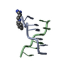
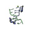
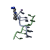
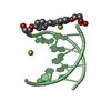
| Entry | Database: PDB / ID: 1ick | ||||||||||||||||||
|---|---|---|---|---|---|---|---|---|---|---|---|---|---|---|---|---|---|---|---|
| Title | LEFT-HANDED Z-DNA HEXAMER DUPLEX D(CGCGCG)2 | ||||||||||||||||||
 Components Components | 5'-D(* Keywords KeywordsDNA / left-handed Z-DNA oligomer | Function / homology | SPERMINE / DNA |  Function and homology information Function and homology informationMethod |  X-RAY DIFFRACTION / SYNCHROTRON / 2DCG model from PDB / Resolution: 0.95 Å X-RAY DIFFRACTION / SYNCHROTRON / 2DCG model from PDB / Resolution: 0.95 Å  Authors AuthorsDauter, Z. / Adamiak, D.A. |  CitationJournal: Acta Crystallogr.,Sect.D / Year: 2001 CitationJournal: Acta Crystallogr.,Sect.D / Year: 2001Title: Anomalous signal of phosphorus used for phasing DNA oligomer: importance of data redundancy. Authors: Dauter, Z. / Adamiak, D.A. History |
|
- Structure visualization
| Structure viewer | Molecule: MolmilJmol/JSmol |
|---|
- Downloads & links
-Download
| PDBx/mmCIF format | 1ick.cif.gz | 26.1 KB | Display | PDBx/mmCIF format |
|---|---|---|---|---|
| PDB format | pdb1ick.ent.gz | 17.7 KB | Display | PDB format |
| PDBx/mmJSON format | 1ick.json.gz | Tree view | PDBx/mmJSON format | |
| Others |  Other downloads Other downloads |
-Validation report
| Arichive directory | https://data.pdbj.org/pub/pdb/validation_reports/ic/1ickftp://data.pdbj.org/pub/pdb/validation_reports/ic/1ick | HTTPS FTP |
|---|
-Related structure data
| Related structure data |  2dcgS S: Starting model for refinement |
|---|---|
| Similar structure data |


-Links
 PDBj
PDBj


- Assembly
| Deposited unit | 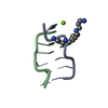
| ||||||||
|---|---|---|---|---|---|---|---|---|---|
| 1 |
| ||||||||
| Unit cell |
|
-Components
| #1: DNA chain | Mass: 1810.205 Da / Num. of mol.: 2 / Source method: obtained synthetically #2: Chemical | ChemComp-SPM / |   Mass: 202.340 Da / Num. of mol.: 1 / Source method: obtained synthetically / Formula: C10H26N4 Mass: 202.340 Da / Num. of mol.: 1 / Source method: obtained synthetically / Formula: C10H26N4#3: Chemical | ChemComp-MG / |   Mass: 24.305 Da / Num. of mol.: 1 / Source method: obtained synthetically / Formula: Mg Mass: 24.305 Da / Num. of mol.: 1 / Source method: obtained synthetically / Formula: Mg#4: Water | ChemComp-HOH / |  Mass: 18.015 Da / Num. of mol.: 88 / Source method: isolated from a natural source / Formula: H2O Mass: 18.015 Da / Num. of mol.: 88 / Source method: isolated from a natural source / Formula: H2O |
|---|
-Experimental details
-Experiment
| Experiment | Method: X-RAY DIFFRACTION / Number of used crystals: 1 |
|---|
- Sample preparation
| Crystal | Density Matthews: 1.74 Å3/Da / Density % sol: 29.13 % | ||||||||||||||||||||||||||||||||||||||||||
|---|---|---|---|---|---|---|---|---|---|---|---|---|---|---|---|---|---|---|---|---|---|---|---|---|---|---|---|---|---|---|---|---|---|---|---|---|---|---|---|---|---|---|---|
| Crystal grow | Temperature: 293 K / Method: vapor diffusion, hanging drop / pH: 7 Details: 30 mM cacodylate buffer, 15 mM MgCl2, 10 mM spermine, 5% MPD, pH 7.0, VAPOR DIFFUSION, HANGING DROP, temperature 293.0K | ||||||||||||||||||||||||||||||||||||||||||
| Components of the solutions |
| ||||||||||||||||||||||||||||||||||||||||||
| Crystal grow | *PLUS | ||||||||||||||||||||||||||||||||||||||||||
| Components of the solutions | *PLUS
|
-Data collection
| Diffraction source | Source: SYNCHROTRON / Site: NSLS  / Beamline: X8C / Wavelength: 0.98 Å / Beamline: X8C / Wavelength: 0.98 Å |
|---|---|
| Detector | Type: ADSC QUANTUM 4 / Detector: CCD / Date: Aug 15, 2000 / Details: mirrors |
| Radiation | Monochromator: Si 111 CHANNEL CUT DOUBLE / Protocol: SINGLE WAVELENGTH / Monochromatic (M) / Laue (L): M / Scattering type: x-ray |
| Radiation wavelength | Wavelength: 0.98 Å / Relative weight: 1 |
| Reflection | Resolution: 0.95→20 Å / Num. all: 16121 / Num. obs: 16121 / % possible obs: 99.8 % / Observed criterion σ(F): -3 / Observed criterion σ(I): -3 / Redundancy: 12 % / Biso Wilson estimate: 4.2 Å2 / Rmerge(I) obs: 0.038 / Rsym value: 0.038 / Net I/σ(I): 58.7 |
| Reflection shell | Resolution: 0.95→0.98 Å / Redundancy: 12 % / Rmerge(I) obs: 0.129 / Mean I/σ(I) obs: 9.7 / Num. unique all: 1550 / Rsym value: 0.129 / % possible all: 98.8 |
| Reflection | *PLUS Num. measured all: 193475 |
| Reflection shell | *PLUS % possible obs: 98.8 % |
- Processing
| Software |
| ||||||||||||||||||
|---|---|---|---|---|---|---|---|---|---|---|---|---|---|---|---|---|---|---|---|
| Refinement | Method to determine structure: 2DCG model from PDB Starting model: 2DCG from PDB Resolution: 0.95→10 Å / Cross valid method: electron density maps / σ(F): -99 / σ(I): -99 / Stereochemistry target values: Parkinson et al. Details: Anisotropic model for all atoms. The average rms error from the inversion of the lsq matrix is 0.01 Angstrom.
| ||||||||||||||||||
| Refinement step | Cycle: LAST / Resolution: 0.95→10 Å
| ||||||||||||||||||
| Refine LS restraints |
| ||||||||||||||||||
| LS refinement shell | Resolution: 0.95→10 Å / Rfactor Rfree error: 0
| ||||||||||||||||||
| Software | *PLUS Name: SHELXL-97 / Classification: refinement | ||||||||||||||||||
| Refinement | *PLUS Num. reflection all: 15782 / Num. reflection obs: 16102 / Rfactor all: 0.0871 | ||||||||||||||||||
| Solvent computation | *PLUS | ||||||||||||||||||
| Displacement parameters | *PLUS | ||||||||||||||||||
| Refine LS restraints | *PLUS
|
